Imaging Methods
- Introduction
- Bias Modulated - Scanning Ion Conductance Microscopy (SICM)
- Scanning Electrochemical Cell Microscopy (SECCM)
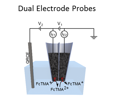
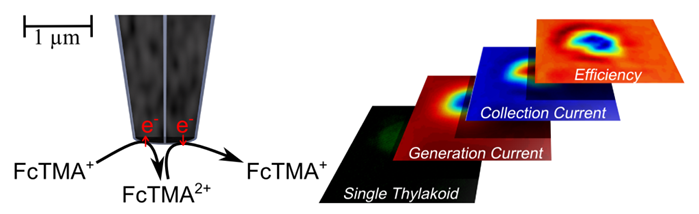
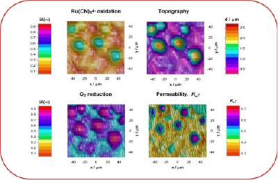
- Scanning Electrochemical Microscopy (SECM)
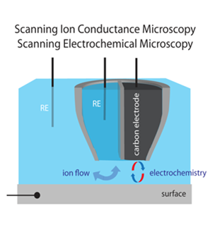
- Combined SICM-SECM
- Biological Systems
- Supported bilayers
- Dentine
- Enamel
- Modelling
IntroductionThe ability to visualise surfaces and interfaces is of critical importance towards understanding dynamic physicochemical processes. The Electrochemistry and Interfaces group at Warwick is well-known for the development of a number of novel electrochemical imaging techniques that have wide-ranging applications in the physical and life sciences. These include scanning electrochemical cell microscopy (SECCM), which is opening up new nanoscale views of electrochemical interfaces, scanning electrochemical microscopy (SECM), new modes of scanning ion conductance microscopy (SICM) and various hybrid techniques, such as combined SECM-SICM and other methods. Further details can be found in the related sections on this page. Central to the diversification of electrochemical imaging methods is the fabrication and characterisation of new electrochemical nanoprobes that offer new capability and functionality. |
   |
|
|
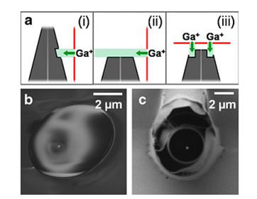
Top: Laser Pulling of Glass Pipettes Middle: Focused Ion Beam milling of tip geometry Bottom: Carbon Filling of Glass Pipettes |
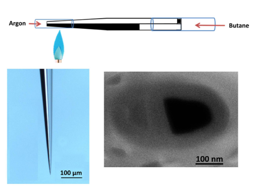
Bias Modulated Scanning Ion Conductance Microscopy (SICM)Scanning ion conductance microscopy (SICM) is regularly used to determine, in a noncontact manner, the topography of a sample. We have developed a new method Bias Modulated scanning Ion Conductance Microscopy (BM-SICM) which, by oscillating the applied bias between the SICM nanopipet probe and the reference electrode in the bulk solution generates a feedback signal to control the distance between the end of a nanopipet and a surface. This development eliminates the need to physically oscillate the probe to generate an oscillating ion current feedback signal, as needed for conventional SICM modes. Moreover, bias modulation allows a feedback signal to be generated without any net ion current flow and phase-detection opens up the mode to the prospect of faster imaging. Bias Modulated Scanning Ion Conductance Microscopy |
|
|
|
|
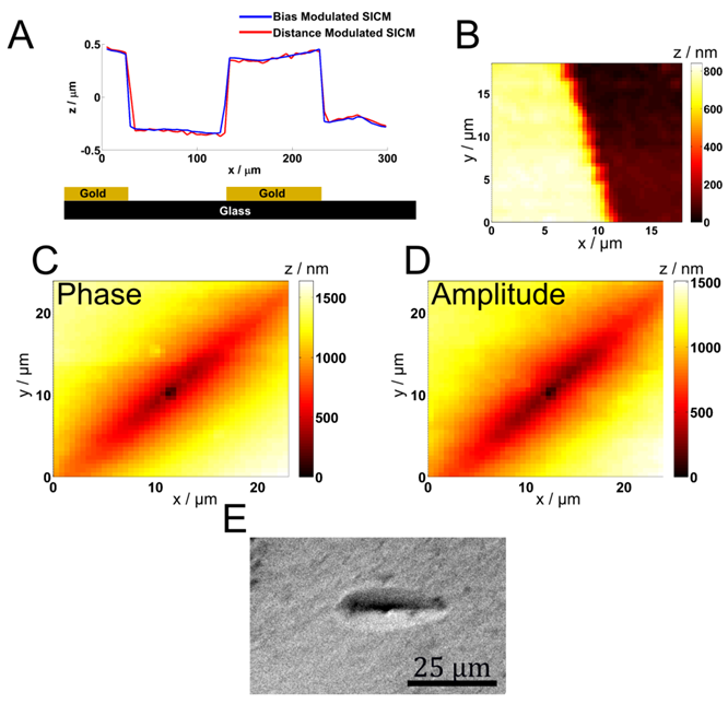 Bias modulated (BM)-SICM is compared to conventional SICM imaging through measurements of substrates with distinct topographical features and yields equivalent results (A and B). Finally, BM-SICM with both amplitude and phase feedback is used for topographical imaging of subtle etch features in a calcite crystal surface(C and D) compared to an SEM immage (E).
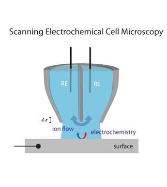
Scanning Electrochemical Cell Microscopy (SECCM)This technique is a highly localized electrochemical measurements and imaging technique using a simple, mobile theta pipet cell. Each channel is filled with electrolyte solution and a Ag/AgCl electrode, between which a bias is applied, resulting in a conductance current across a thin meniscus of solution at the end of the pipet, which is typically deployed in air or a controlled gaseous environment. When the pipet is oscillated, an oscillating component in the conductance current is generated. This oscillating current component can be used to maintain gentle contact of the solution from the pipet cell with the surface and as a set point for high resolution topographical imaging with the pipet. Simultaneously, the mean conductance current that flows between the pipet channels can be measured and is sensitive to the local nature of the interface, informing one, for example, on wettability and ion flow into or out of the surface investigated. Conductor or semiconductor surfaces can be connected as a working electrode, with one of the electrodes in the pipet serving as a quasi-reference electrode. This pipet cell then constitutes part of a dynamic electrochemical cell, with which direct voltammetric−amperometric imaging can be carried out simultaneously with conductance and topographical imaging. This provides multifunctional electrochemical maps of surfaces and interfaces at high spatial resolution. Localized High Resolution Electrochemistry and Multifunctional Imaging: Scanning Electrochemical Cell Microscopy |
|
 |
Scanning Electrochemical Microscopy (SECM) |
  |
 |
|
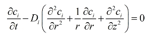
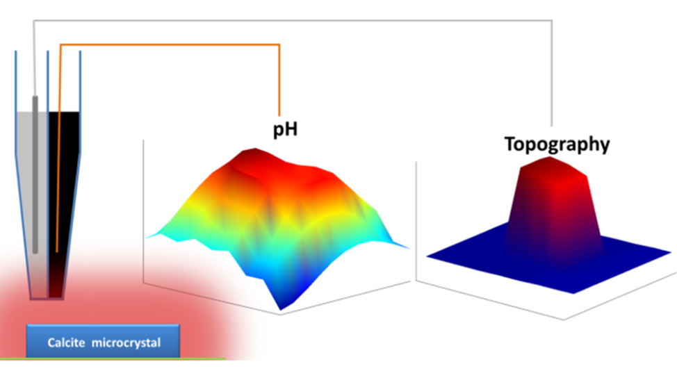
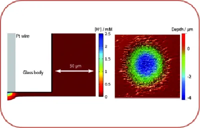
Combigned SICM-SECMThe easy and cheep fabrication and use of nanoscale dual function pH-scanning ion conductance microscopy (SICM) probes has been developed here at Warwick. One Example of this type of probe incorporate an iridium oxide coated carbon electrode for pH measurement and an SICM barrel for distance control, enabling simultaneous pH and topography mapping. The fabricated probes, with pH and SICM sensing elements are typically on the 100 nm scale. The capability of the pH-SICM probe was demonstrated by detecting both pH and topographical changes during the dissolution of a calcite microcrystal in aqueous solution. This system illustrates the quantitative nature of pH-SICM imaging, because the dissolution process changes the crystal height and interfacial pH (compared to bulk), and each is sensitive to the rate. Fabrication and Characterization of Dual Function Nanoscale pH-Scanning Ion Conductance Microscopy (SICM) Probes for High Resolution pH Mapping B. P. Nadappuram, K. McKelvey, R. Al-Botros, A. W. Colburn, and P. R. Unwin, Anal. Chem., 2013, 85(17), 8070–8074. |
 |
 |
|
|
|
Scanning electrochemical microscopy is widely used to investigate various aspects of biological systems such as morphology, metabolism and transport of different signalling molecules. Our research mainly focusses on electrochemical probing of cell surfaces, development of integrated electrochemical microscopy techniques capable of monitoring and investigating specific metabolites and biological reactions with high spatial resolution and investigation of material transport across biological membranes. A broad range of electrochemical scanned probe techniques are used including SECM, SICM, combined SECM-SICM, IC-SECM and SECCM, These scanning electrochemical microscopy techniques were also integrated with optical microscopy systems such as ultra-fast confocal laser scanning microscopy and other related technologies, many of which are pioneered in our group. |
|
|
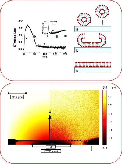
Supported lipid bilayer systems were extensively used as model cellular membranes for the fundamental studies of biophysical processes. A key aspect of our research in this area is to probe and confirm formation of supported lipid bilayer and subsequently study dynamic processes taking place on and across this membrane. Transport of molecules across cell membrane is fundamental processes in cellular biology. They are also becoming increasingly important in many medical, pharmaceutical, and environmental technologies. Our research mainly focusses on development of new method to investigate the formation of supported bilayers on various solid substrate systems by using techniques like AFM, evanescent wave cavity ring down spectroscopy and quantitatively measure the permeation of various molecules via microelectrochemistry, combined with laser confocal scanning microscopy (LCSM) and supported by finite element modeling (FEM).
|
|
|
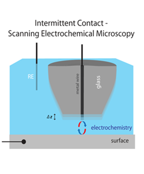
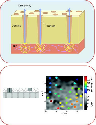
Dentinal hypersensitivity is a common problem, affecting much of the adult population. It is caused by the movement of fluid through exposed dentinal tubules. Scanning Electrochemical Microscopy (SECM) has been shown to be a powerful technique for the measurement of local solution velocities through human dentine slices, in-vitro, and for assessing quantitatively the effect of treatments on fluid flow. The main goal of our research is to improve the spatial resolution and contrast of SECM measurements of transport through model porous materials by using improved electrochemical techniques like intermittent contact (IC)-SECM, which will open a new way to understand and treat dentinal hypersensitivity.
|
|
|
The rate of dental enamel dissolution is very important in the area of dental science in the context of acid erosion and in understanding how this process can be inhibited. In our group, we use scanning electrochemical microscopy (SECM) for the quantitative study of acid-induced bovine enamel dissolution. High rates of mass transport in SECM allow the surface kinetics of dissolution to be evaluated. SECM is able to deliver high, controllable, and local acid challenges in a defined way and that multiple dissolution measurements can be performed on one sample, eliminating intersample variability effects.
|
|
|
Finite element simulations are used to provide a theoretical framework for experiments that are too complicated to approach analytically. A simulation domain is defined with boundary conditions and internal physics based upon the experiment to be modelled. The FEM provides a means of approximating the continuous space within the domain to a finite number of points (elements). At each element the equations specified by the user are solved to provide a solution. For example Fick's 2nd Law of diffusion, in 2D-axisymmetrical co-ordinates are defined as:
In addition to providing numerical data Comsol Multiphysics can provide visual representations which are valuable for understanding the key events for understanding experimental results, or designing experiments. For example the expansion of the diffusion layer of a mediator species at a macro disc electrode when the potential is swept and the corresponding linear sweep voltammogram are shown in the figure. |