
Imaging Team
Electrochemical Imaging Team
WEIG's Imaging Team, led by Prof. Pat Unwin, investigates electrochemical phenomena at the nanoscale.
We use primarily scanning electrochemical probe microscopy techniques to investigate electrode surfaces, membranes, nanoparticles, biological material, etc., and conduct non-invasive analysis, under a range of conditions.

Developing smart instrumentation for operando monitoring of electrochemical processes
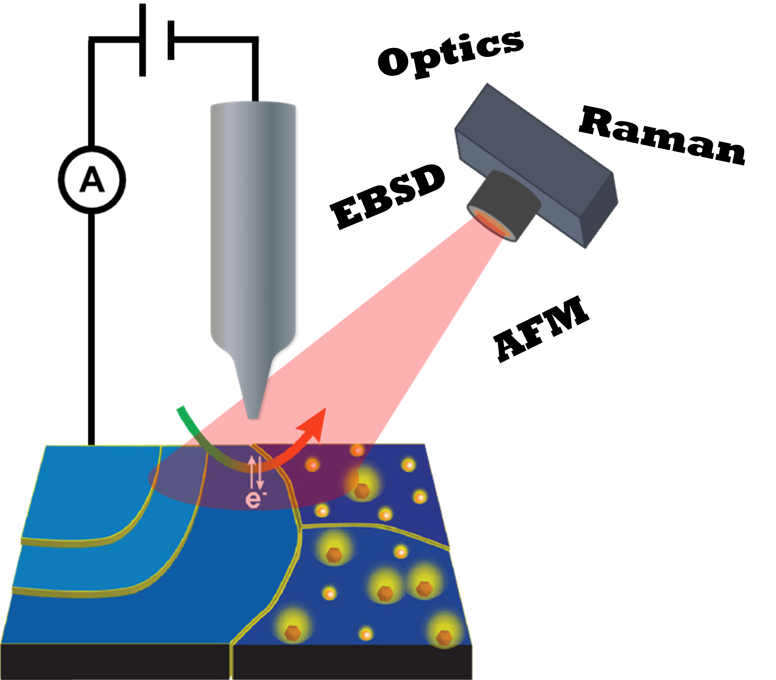
Electrochemical probes and correlative microscopies
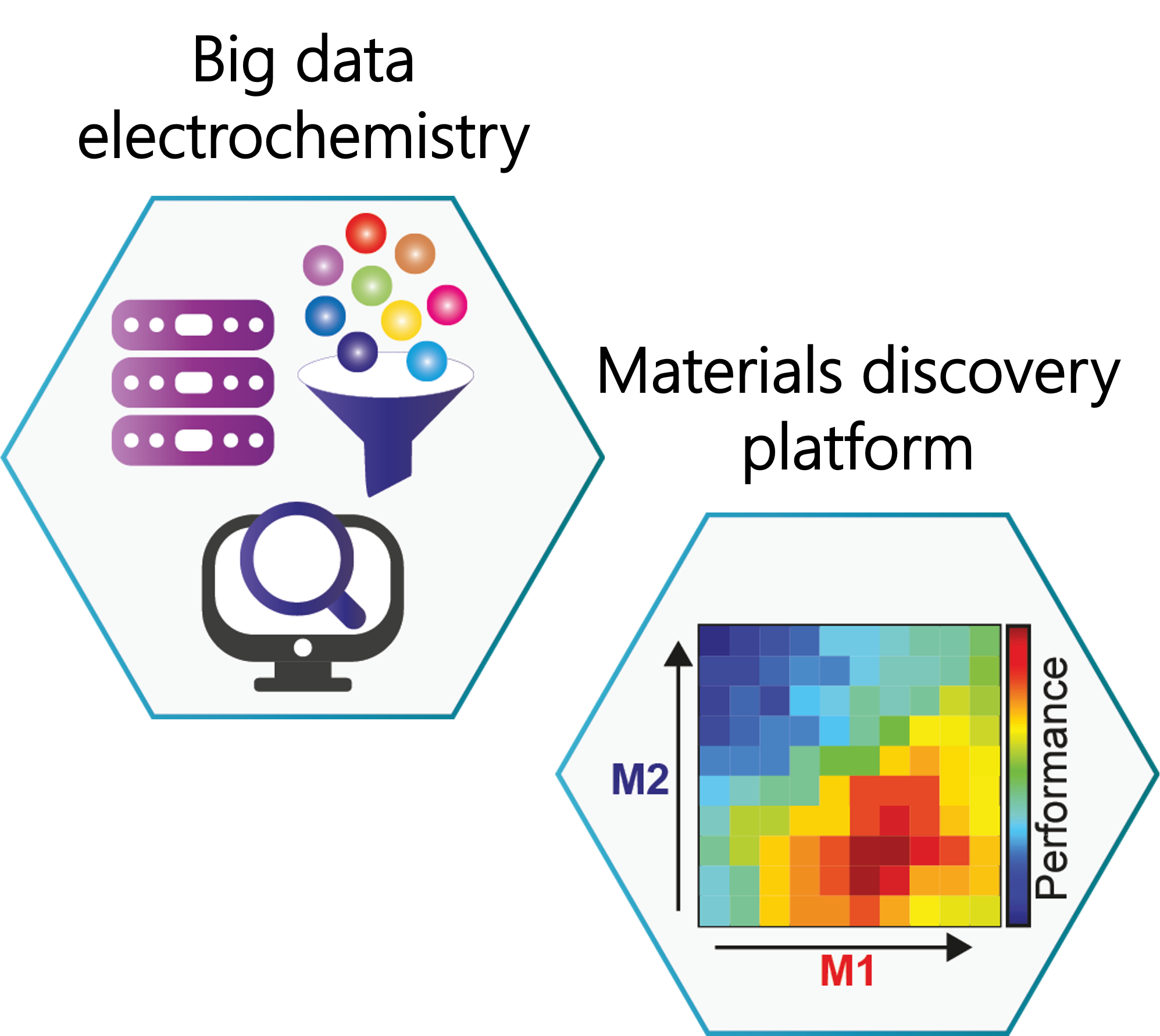
Materials discovery through big data electrochemistry

O. J. Wahab, E. Daviddi, et al., Nature, 2023, 620, 782-786
https://doi.org/10.1038/s41586-023-06247-6Link opens in a new window
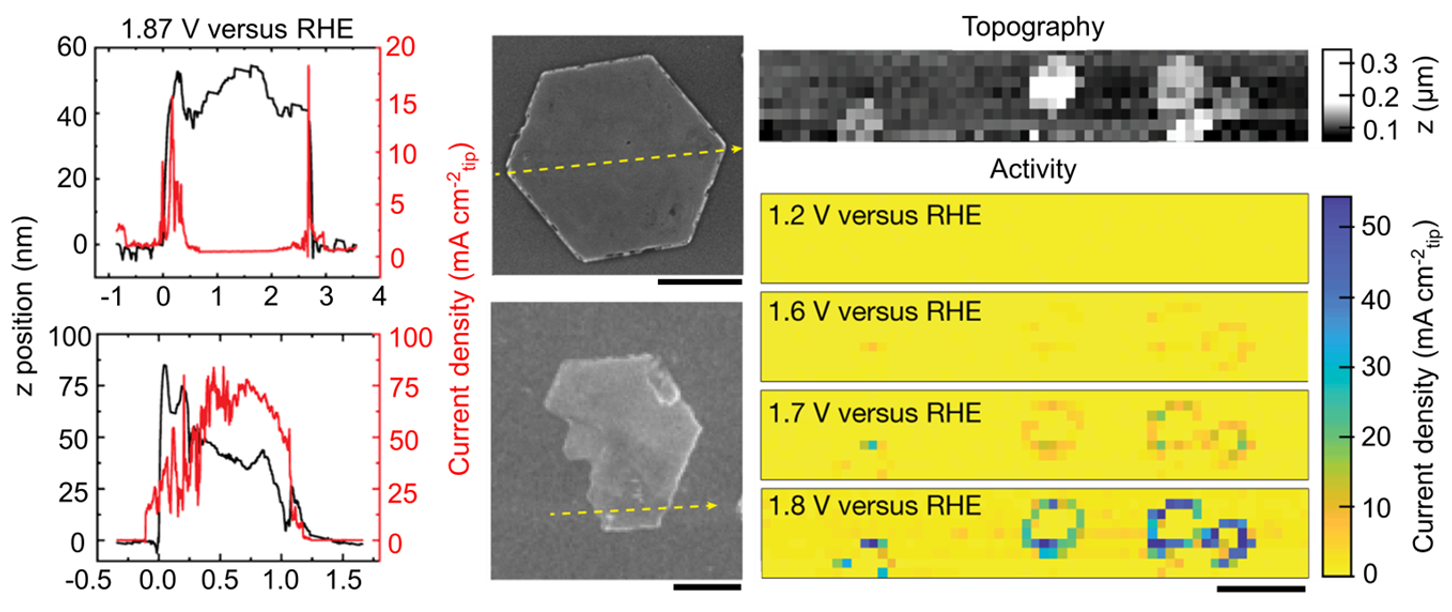
Mapping the local electrochemical activity at
two-dimensional materials with nanoscale resolution
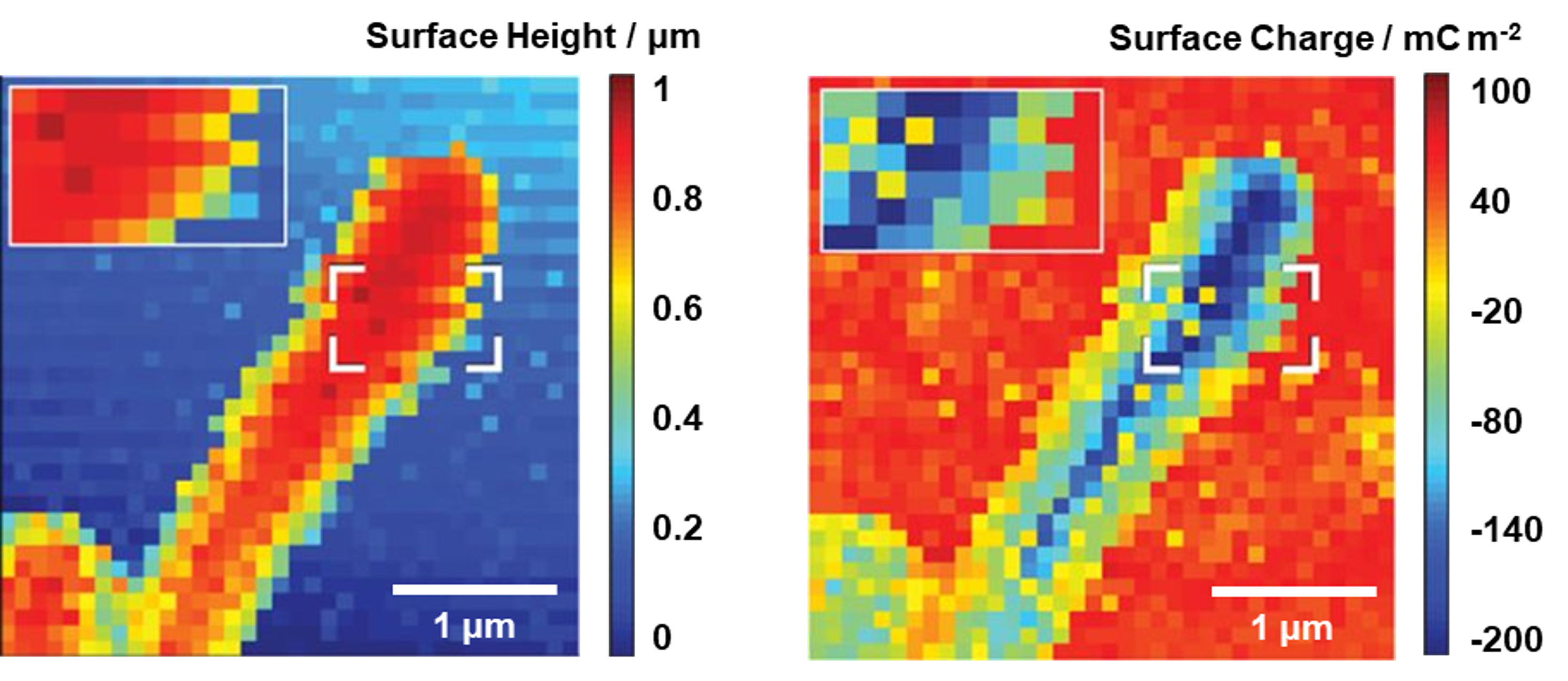
A miniature droplet electrochemical cell facilitates the investigation of the local surface activity. In this work we aimed to determine if protons can pass through one-atom-thick graphene and hexagonal boron nitride membranes. A measureable current was correlated with nanoscale wrinkles and stress areas on the membranes (as visualised with atomic force microscopy adhesion maps).
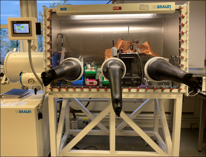
Intelligent scanning electrochemical probes for high-throughput single-entity measurements
We perform scanning electrochemical cell microscopy (SECCM) experiments inside a glovebox to study battery materials under industry-relevant conditions. An optics-guided, intelligent scanning protocol enables the investigation of multiple targets at a fraction of the standard experiment time.
E. B. Tetteh, D. Valavanis, et al., Angew. Chem. Int. Ed., 2023, 62 (9), e202214493
https://doi.org/10.1002/anie.202214493Link opens in a new window
D. Martín-Yerga, D. C. Milan, X. Xu, et al., Angew. Chem. Int. Ed., 2022, 61 (34), e202207184
https://doi.org/10.1002/anie.202207184Link opens in a new window
Correlative multi-microscopy for catalysis
SECCM provides simultaneous topography and electrochemical activity, reaching sub-particle-level resolution. Here, we revealed the dominant active sites of β-Co(OH)2 particles for oxygen evolution reaction.
J. T. Mefford, A. R. Akbashev, M. Kang, et al. Nature, 2021, 593, 67–73
https://doi.org/10.1038/s41586-021-03454-xLink opens in a new window
Multi-functional imaging of cells, bacteria, and other biological samples
Electrochemical probes can get close to, inspect, and manipulate, many types of live biological samples. In this work, a glass nanopipette probe was used to simultaneously measure the topography and surface charge of E. coli bacteria.
K. Cremin, B. A. Jones, et al., Anal. Chem., 2020, 92 (24), 16024-16032
https://doi.org/10.1021/acs.analchem.0c03653Link opens in a new window
To view more information on the scanning electrochemical probe microscopy techniques that we utilise, or some of the systems that we study, you can visit the sub-pages Imaging Methods or Crystallisation.