
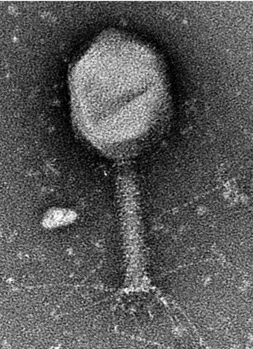
How to catch a virus
Step 1
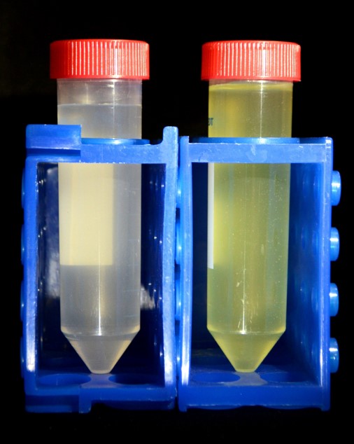
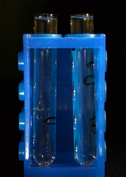
Here we have two water samples for testing. The left is from a river, the right from a pond. Even thought they look quite different they contain roughly the same sort of things - bits of dirt, algae and other microorganisms.
Step 2
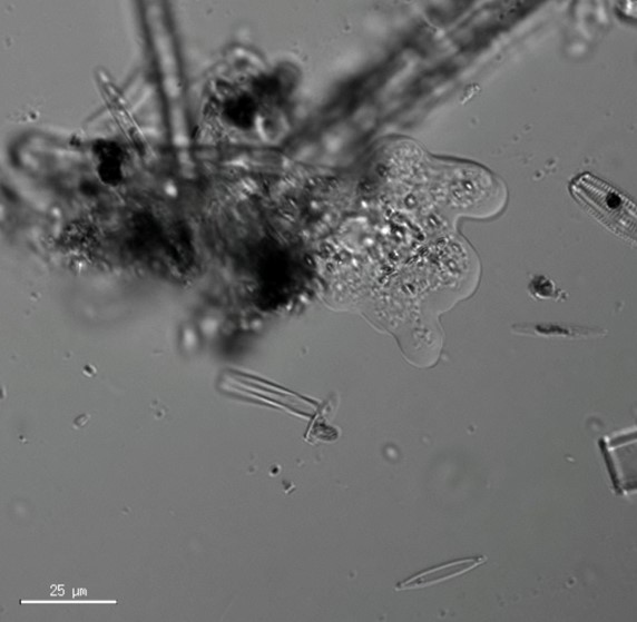
This is an amoeba, they may look great under the microscope but they’re not the part we’re interested in. They’re microscopic but they’re far too big for the electron microscope. We need to get all those microbes and other cloudy stuff out of the water.
Step 3
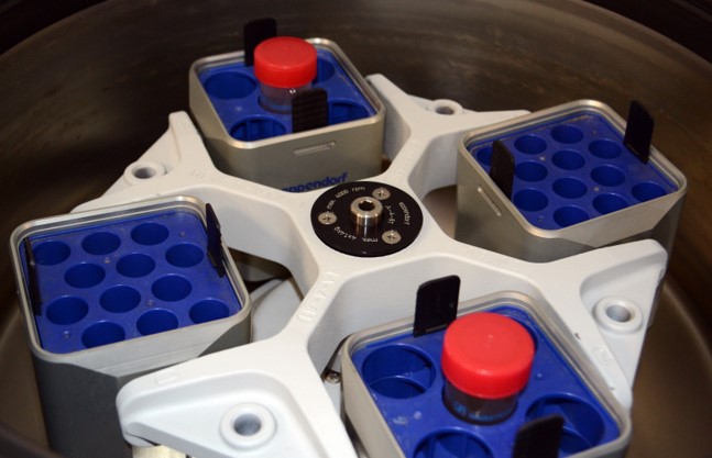
We could just let gravity do the work – if we leave it to sit then most of the stuff will settle to the bottom of the tube but it’ll take ages and we don’t want to wait. We can give gravity a helping hand and speed things up if we put our tubes into a centrifuge and spin them at high speed.
Step 4
At 3000 RPM we’re subjecting the tubes to 6000x the force of gravity. At this speed after just 10 minutes, all the bits of sediment, bacteria and other things are pulled to the bottom of the tube as a pellet.
What we need is the liquid above the pellet - the supernatant. It looks as clear as tap water but you wouldn’t want to drink it – it’s full of loads of things that were too small to get trapped by the first spin and that includes the viruses we’re interested in.
Step 5
We’ll need to spin the sample again to get to our viruses – but a lot faster than last time. So fast that we need special titanium buckets to protect the tubes from the extreme forces.
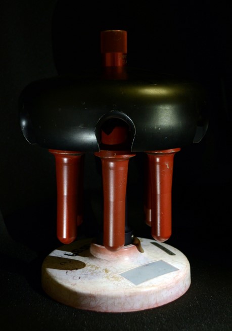
Step 6
Special titanium buckets
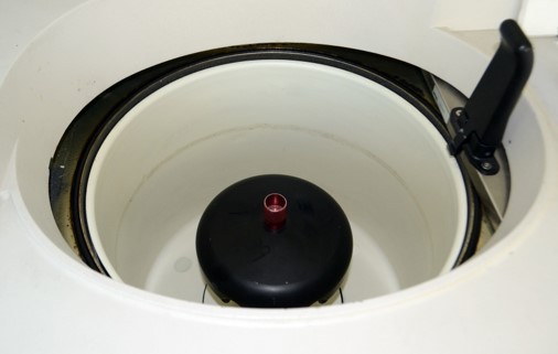
Step 7
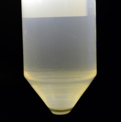
The ultracentrifuge spins our samples at 40,000 RPM – at that speed they’re feeling around 250,000 times the force of gravity. At that speed even tiny things like viruses get forced out of solution and dragged to the bottom of the tube to make a pellet of yellow goo.
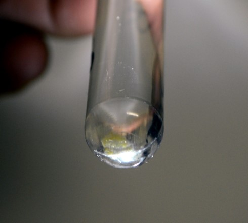
Step 8
Yellow goo at the bottom of the tube
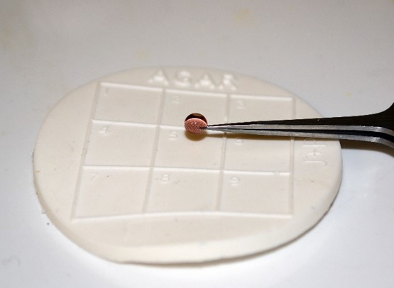
Step 9
Just one hundredth of a millilitre of sample is all we need. The viruses should stick as we blot away the water with a piece of filter paper. The sample gets stained with a solution of heavy metal compounds to make it easier to see in the microscope. Then it's put into the sample holder, loaded into the microscope and the fun begins!
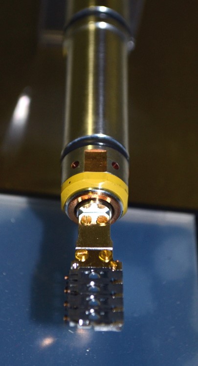
Step 10
Put the sample in the sample holder
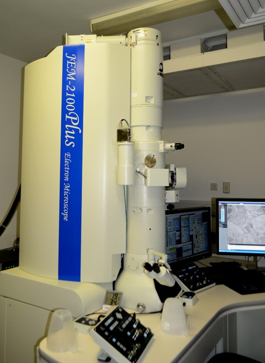
Step 11
The microscope we use to image the samples. It uses electrons!
Step 12
Look at the sample!