W-CAS Older News
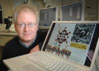
Imaging the Structure, Symmetry, and Surface-Inhibited Rotation of Polyoxometalate Ions on Graphene Oxide
Jeremy Sloan†, Zheng Liu‡, Kazu Suenaga‡, Neil R. Wilson†, Priyanka A. Pandey†, Laura M. Perkins§, Jonathan P. Rourke, and Ian J. Shannon§ † Department of Physics, University of Warwick, Coventry, CV4 7AL, U.K. Nano Letters, October 26th 2010. |
Atomic-resolution imaging of discrete [γ-SiW10O36]8− lacunary Keggin ions dispersed onto monolayer graphene oxide (GO) films by low voltage aberration corrected transmission electron microscopy is described. Under low electron beam dose, individual anions remain stationary for long enough that a variety of projections can be observed and structural information extracted with ca. ±0.03 nm precision. Unambiguous assignment of the orientation of individual ions with respect to the point symmetry elements can be determined. The C2v symmetry [γ-SiW10O36]8− ion was imaged along its 2-fold C2 axis or orthogonally with respect to one of two nonequivalent mirror planes (i.e., σv). Continued electron beam exposure of a second ion imaged orthogonal to σv causes it to translate and/or rotate in an inhibited fashion so that the ion can be viewed in different relative orientations. The inhibited surface motion of the anion, which is in response to H-bonding-type interactions, reveals an important new property for GO in that it demonstrably behaves as a chemically modified (i.e., rather than chemically neutral) surface in electron microscopy. This behavior indicates that GO has more in common with substrates used in imaging techniques such as atomic force microscopy and scanning tunneling microscopy, and this clearly sets it apart from other support films used in transmission electron microscopy. |
Localized High Resolution Electrochemistry and Multifunctional Imaging: Scanning Electrochemical Cell Microscopy
|
|
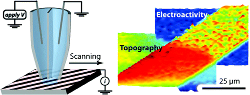
We describe highly localized electrochemical measurements and imaging using a simple, mobile theta pipet cell. Each channel (diameter <500 nm) of a tapered theta pipet is filled with electrolyte solution and a Ag/AgCl electrode, between which a bias is applied, resulting in a conductance current across a thin meniscus of solution at the end of the pipet, which is typically deployed in air or a controlled gaseous environment.
|
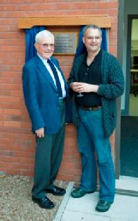
Opening of the newly built FTICR Laboratory by Professor Fred McLafferty of Cornell University
 |
Thursday 9 September saw the official opening of the newly built Ion Cyclotron Resonance Laboratory at Millburn House, which houses state-of-the-art Fourier transform ion cyclotron resonance (FTICR) mass spectrometers.
Opening in pictures here! |


