
Linear Dichroism (LD)
Linear dichroism (LD) is the difference in absorption, A, of light linearly polarized parallel (//) and perpendicular (⊥) to an orientation axis:
LD=A//–A⊥
LD is used with systems that are either intrinsically oriented or are oriented during the experiment. The signal that is measured contains information about the orientation of sub-units of the system relative to the orientation axis.
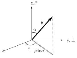
The orientations of an electric dipole transition moment, μ, (whose length/magnitude is μ). The magnitude of the projection of μ onto the z axis is μcosα and onto the x/y plane is m sina. The y component of this projection is μsinαsinγ. For a perfectly oriented molecular system Z = z and S = 1.
How does it work?
LD is related to the more commonly used technique of circular dichroism (CD), in that both require the difference between the absorbances of different polarized light beams to be measured and CD spectropolarimeters can be adapted to produce the required alternating beams of polarized light for LD. Interpretation of LD data is based on the figure above. For the standard geometry, if α = 0° (i.e. the transition of interest is oriented parallel to the orientation direction) then LD>0; conversely, if α = 90° (i.e. the transition of interest is oriented perpendicular to the orientation direction) then LD<0; and LD=0 when α= 54.7°, the so-called magic angle. With biomacromolecules, we often know the details of the spectroscopy of the chromophores (such as nucleic acid bases or amino acids) so the LD immediately gives us qualitative orientation information. Quantitative analysis is possible.
Applications:
Biological structure analysis, particularly DNA, RNA, fibrous proteins, membrane proteins, lipid structures, carbon nanotibes. Kinetics of such systems. Ligand binding (only those small molecules actually bound to an oriented macromolecule show an LD signal)
Sample handling requirements:
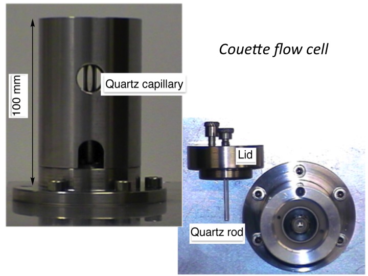
Usually solutions which have a maximum absorbance of 1 in the region of interest. Buffer or other matrix components should not have a significant absorbance. Typical concentrations range from 0.1–1 mg/mL for a 0.5 mm path length Couette flow cell.
Complementary techniques:
Absorbance Spectroscopy; Circular Dichroism
Warwick expertise:
6 CD machines, 3 of which will measure linear dichroism. Jasco J-815 (with fluorescence detected spectroscopy, multiple sample holder, and temperature control) , Jasco J-815, Jasco J-J720, Jasco J-600, 2 Biologic MOS-450 stopped flow spectrometers. Warwick has developed the only commercially available Couette flow cells for LD which are available from Dioptica Scientific Ltd.
Contact:
Claire Gerard: / 07385 145064