
Raman Microscopy
A Raman microscope enables spectra to be measured from defined points (down to around 5 μm) on a sample. Applications include phase identification, strain studies and detection of impurities. Mapping studies can be carried out, with spectra collected over a defined array, allowing the location and identifications of different components in a complex material.
How does it work?
When light is scattered from a crystal, solution or molecule, most photons are elastically scattered. The scattered photons have the same energy (frequency) and, therefore, wavelength, as the incident photons. However, a small fraction of light (approximately 1 in 107 photons) is scattered at optical frequencies different from the frequency of the incident photons. The process leading to this inelastic scatter is termed the Raman effect. Raman scattering can occur with a change in vibrational, rotational or electronic energy of the scatterer.
If the substance being studied is illuminated by monochromatic light, for example from a laser, the spectrum of the scattered light consists of a strong line (the exciting line) of the same frequency as the incident illumination together with weaker lines on either side shifted from the strong line by frequencies ranging from a few to about 3500 cm-1. The lines of energy (frequency) less than the exciting photons are called Stokes lines, those at higher energy are called anti-Stokes lines. Typically it is the Stokes lines which are recorded in a Raman spectrum. The combination of a spectrometer with a microscope allows a precise point on the surface of a sample to be selected for analysis.
Applications:
Non destructive analysis of paint pigments and pottery; cell research; disease detection; drug design and pharmaceutical materials; characterisation of drug-cell interactions; microbiology and cell sorting; cosmetics and in vivo skin analysis; characterisation and polymorphy of pharmaceuticals; physical and chemical quantitative behaviour prediction in polymers; strain measurements in thin film semiconductor layers; observation of oxidation kinetics on rusting surfaces.
Sample Handling Requirements:
Solids, liquids, gases (in general not suitable for metals or their alloys).
Complementary Techniques:
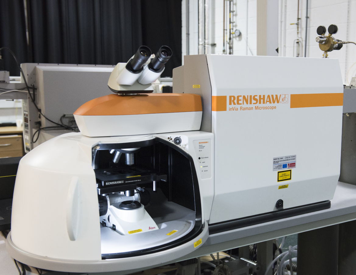
Warwick Capability:
Renishaw Invia Reflex. Excitation available at 325, 442, 514, 633 and 785 nm wavelengths, and temperatures from 4 K to 600 K
Biotools Chiral RAMAN - Contact i.hancox@warwick.ac.uk for further information.
Contact:
Claire Gerard: / 07385 145064
Professor Mark Newton, / 024 76 574 358.