
MSc Project 3
Combined Scanning Electrochemical Microscopy - Scanning Ion Conductance Microscopy (SECM-SICM)
Imaging and Probing Water and Ion Transport at the Nanoscale
Supervised by Prof. Pat Unwin
Background
High-resolution electrochemical microscopy techniques have become increasingly attractive as a means of mapping the function and topography at surfaces and interfaces. Such techniques have proven useful for interpreting a wide range of biological and chemical systems, from the adsorption and transport of species across cell membranes,1 to the dissolution and growth processes of materials such as ionic crystals.2 Scanning electrochemical microscopy (SECM) has developed as the prevailing technique to quantitatively visualise interfacial functional processes.3 However, the main drawback of SECM is that it lacks distance (feedback) control, which makes it difficult to distinguish whether an observed change in current is due to a functional or structural attribute. Variants of SECM have been developed to combat this problem including SECM-atomic force microscopy (AFM),4 and intermittent contact (IC)-SECM.5 Scanning ion conductance microscopy (SICM) is another electrochemical microscopy technique that uses similar technologies to SECM, but measures the ionic conductance between two electrodes.6 Combining SECM and SICM (SECM-SICM) would provide a powerful new imaging technique, utilising the feedback control of SICM to map out topography, whilst simultaneously using SECM to scan for chemical activity on the surface.
In my project, I developed a method to fabricate combined SECM-SICM tips and used them to image, with nanoscale resolution, the surface of model conducting/insulating substrates (Fig. 1) and maize root cells.
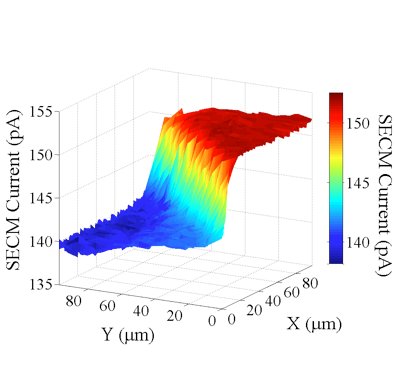 Fig. 1: SECM Current as the SECM-SICM probe scans over the edge of a conducting gold band (red region) and an insulating glass substrate (blue region).
References
[1] Matsue, T.; Shiku, H.; Yamada, H.; Uchida (1994) Permselectivity of voltage-gated alamethicin ion channel studied by microamperometry. J Phys Chem 98: 11001
[2] Macpherson, J. V.; Unwin, P. R.; Hillier, A. C.; Bard, A. J. (1996) In-situ imaging of ionic crystal dissolution using an integrated electrochemical/AFM probe. J Am Chem Soc 118: 6445
[3] Edwards, M. A.; Martin, S.; Whitworth, A. L.; Macpherson, J. V.; Unwin, P. R. (2006) Scanning electrochemical microscopy: principles and applications to biophysical systems. Physiol Meas 27: R63
[4] Macpherson, J. V.; Unwin, P. R. (2000) Combined scanning electrochemical-atomic force microscopy. Anal Chem 72: 276
[5] McKelvey, K.; Edwards, M. A.; Unwin, P. R. (2010) Intermittent Contact- Scanning Electrochemical Microscopy (IC- SECM): A New Approach for Tip Positioning and Simultaneous Imaging of Interfacial Topography and Activity. Anal Chem 82: 6334-6337
[6] Hansma, P.; Drake, B.; Marti, O.; Gould, S.; Prater, C. (1989) The scanning ion-conductance microscope. Science 243: 641