
LIPSyM for Cell Detection
Abstract
Diabetes can be associated with a reduction in functional β cell mass, which must be restored if the disease is to be cured or progress is to be arrested. To study the β cell count, it is necessary to also determine the number of nuclei within the insulin stained area. It can take a single experimentalist several months to complete a single study of this kind, results of which may still be quite subjective. In this paper, we propose a framework based on a novel measure of local symmetry for detection of β cells. The local isotropic phase symmetry measure (LIPSyM) is designed to give high values at or near the cell centers. We demonstrate the effectiveness of our algorithm for detection of two types of specific cells in histology images, β cells in mouse pancreatic sections and lymphocytes in human breast tissue. Experimental results for these two problems show that our algorithm performs better than human experts for the former problem, and outperforms the best reported results for the latter.
Publication
- Kuse M., Wang YF., Kalasannavar V., Khan M., Rajpoot N. "Local Isotropic Phase Symmetry Measure for Detection of Beta Cells and Lymphocytes," Journal of Pathology Informatics, 2:2, 2011 [DOI]
An earlier version of this paper was presented at the MICCAI workshop on Histology Image Analysis (HIMA), held in Toronto, in Sep 2011.
Biological Background
Glucose homeostasis depends on the release of insulin and glucagon by β cells and α cells within the pancreatic islets of Langerhans, which respectively increase or reduce glucose disposal in peripheral tissues. All major types of diabetes are associated with a reduction in functional β cell mass, which must be restored if the disease is to be cured or progress arrested. Increasingly, drugs are being introduced into clinical practice which may be able to arrest or potentially in the future even reverse this decline at least in Type 2 Diabetes. Not surprisingly, much research in the academic and private sectors is devoted to functional testing of genetic manipulations and therapeutic strategies aimed at interfering with loss of beta cell mass or that might restore or expand endogenous beta cell mass in animal models. Moreover, increasingly post mortem analysis of β and islet cell mass are undertaken in patients. It is not currently possible to measure changes in β cell or even islet mass during life as no sufficiently sensitive or specific imaging tools exist. Therefore, all depends on an accurate assessment of beta cell mass post mortem. In all such cases it is necessary to estimate β cell mass by immunohistochemical (IHC) staining of pancreas tissue sections and then, for at least 5-10 levels, to measure the area of insulin interactivity and divide this by the total pancreas area (mass can then be assessed by multiplying the ratio by the pancreas weight). To assess β cell count, it is necessary to also determine the number of nuclei within the insulin stained area.
Motivation and Objectives of LIPSyM
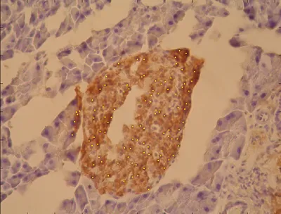
Normal practice to obtain the beta cell count is that, a human expert counts the number of β cell nuclei from digital images obtained from a whole slide scanner. Obtaining the count of β cells in such a way is very labor intensive and subject to observer bias due to massive image sizes and subjectivity in human judgment. Many hours of microscopy and image capture are required followed by very time consuming analysis. Thus, it is not unusual for a single experimentalist to take many months to complete a single study of this kind. To ease this laborious and intensive work of a human expert, we have developed a method for automatic annotation of β cells from H-DAB stained images. The method was finally converted to a software. This software can do automatic detection very quickly over a large number of images. An option was provided for the human expert to alter the auto-annotation result. The software generated reports containing the co-ordinates and the count of β cells for each of the provided input.
This software is currently being used at the Life Science Department, University of Warwick, UK. The code for experimentation and the software are available for download from this site.
About the Method - LIPSyM
We propose a framework based on a novel measure of local symmetry for detection of β cells. The local isotropic phase symmetry measure (LIPSyM) is designed to give high values at or near the points of symmetry. Since, the cells are circular or elliptical in shape, they exhibit high levels of symmetry. This very characteristic observation is the basis of proposed method. More details on the measure can be found in the related publication titled “Local Isotropic Phase Symmetry based Algorithm for Detection of β Cells”.
Results obtained with LIPSyM
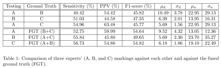
The evaluation of LIPSyM method for β cell detection was done by comparing the results with fused annotations by three independent human experts. Also, LIPSyM was compaired with another simple way of cell detection – Laplacian of Gaussian (LOG). Also, comparison was made between the annotations of different human experts. The results are as tabulated in table below. The details of evaluations of can be found in the related publication.

Downloads
Warwick Beta Cell Dataset
We invite fellow researches to download our dataset for development of further robust and high performing methods. The dataset contains 20 Hematoxylin+DAB stained images. Along with the H+DAB stained images we are also providing markings by three independent human experts and their fused markings. Also, markings by LIPSyM are provided for comparison.
Click here to start the download.
MATLAB Code
Please email Manohar Kuse or Nasir Rajpoot for the latest release of the MATLAB code for LIPSyM.
Disclaimer
This software is free only for non-commercial use. It must not be distributed without prior permission of the author. We are not responsible for implications from the use of this software.