
Lithography/ 3d Imaging
Lithography
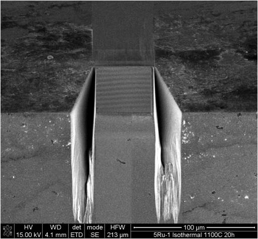
The Jeol 4500 FIB/SEM is not only capable of imaging samples by SEM, but is also equiped with a Ga+ ion source to allow precise focused ion beam (FIB) cutting of a sample. This can be used to make complex structures such as that below:
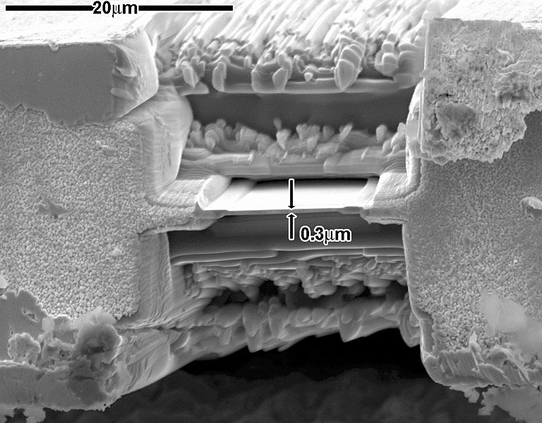 |
Sample modified by FIB/SEM |
3d Imaging
By sequentially imaging and removing areas using the ion beam on the Jeol 4500 FIB/SEM, it is possible to build a 3d model of a microstructure. This gives additional information about the material that sectioning may not be able to provide. Areas as large as a 100 micron wide cube can be imaged in this manner.
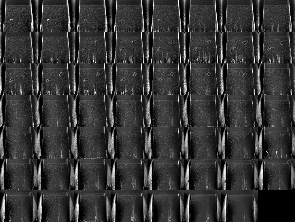
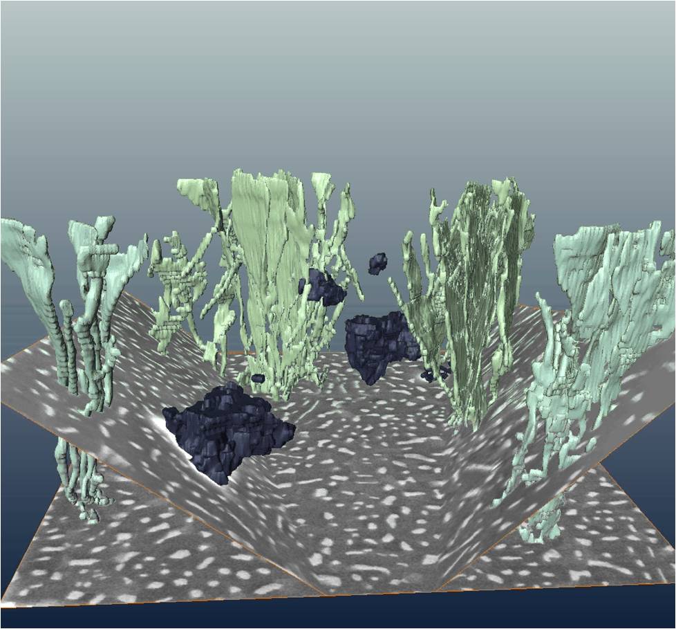
Below are pictures of a specimen that has been 3d imaged. This is a coating on a Ni superalloy turbine blade, which contains Ru-rich precipitates. Sections show the precipitates as bright dots or lines; combining the images into a 3D dataset shows that they actually form a branched structure of parallel sheets, very similar to that of a coral.
 |
 |
 |
Front slice |
Montage of slices |
Reconstruction of 3d volume |