Trace Metals in Medicine Laboratory
Academic responsible - Prof Joanna Collingwood
Our primary focus is imaging and quantifying transition metal ion distribution in the human brain, supporting progress in early detection and diagnosis for neurodegenerative disorders. We use a variety of analytical techniques, including high resolution MRI, synchrotron X-ray microfocus spectroscopy, and magnetometry to characterize the distribution and form of trace metals in tissues and protein aggregates. Please see our recent publications, most of which are open-access.
Contents
Trace Metals in Medicine Laboratory, equipped for sample preparation and analysis. Resources include:
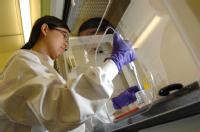
- Wet-lab with chemical safety cabinets, refrigerated centrifuge, balances, ultrapure water supply, and refrigeration equipment.
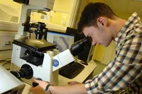
- Containment level 1 area with Class II biosafety cabinets, cryomicrotome sectioning for trace metals analysis in tissues, ultramicrotome sectioning, fluorescence light microscopy (inverted microscopy), and freeze drying.
- Cell culture resources (including incubator and fluorescence plate reader).
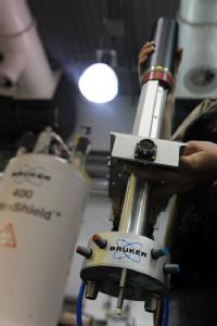
- MRI Probe (for imaging at 9.4 T): we have a state-of-the-art MR microscopy probe that utilizes the 400 MHz spectrometer in Millburn House, under the Science Cities Translational Medicine 2 Programme. It is presently configured for in-vitro work, and the coils are primarily volume coils for 1H imaging; we also have a surface coil, and a 19F volume coil for small samples (<5mm) .
Metal-ion-based neurodegeneration: enabling techniques for understanding, detection and treatment.
Many diseases of the human brain lead, over time, to degeneration of tissue and loss of function. By the time the disease is detected in an individual because of loss of function (whether cognitive or physical), extensive degeneration has in many instances already taken place. Reversing this degeneration presents an enormous challenge; the goal of this project is instead to focus on understanding factors that contribute to causing the degeneration, and to find ways of identifying the degeneration at an early stage in order to i) improve detection, and ii) offer new targets for effective treatment.
A common theme linking many neurodegenerative diseases such as Alzheimer's, Parkinson's, Huntington's, Motor Neurone Disease, and Multiple System Atrophy, is that changes in the regulation of certain trace metals, and/or the proteins responsible for binding and utilizing these metal elements, are apparent. This can include accumulation of certain elements, such as iron, in specific regions of the brain. Our hypothesis is that these changes are disease-specific, and if better understood, may provide windows of opportunity for improved detection and treatment.
Limiting factors affecting present work in this area include:
i) the challenge of extrapolating findings from simple experiments in the laboratory to the complexity of the biochemical environment in the brain;
ii) the challenge of accurate sensitive detection of trace metal elements in the brain - both for measurement in the living brain using clinical techniques, and for laboratory analysis of brain tissue.
In this research, we use experimental and computational approaches to describe, predict, and test evidence of altered trace metal regulation in neurodegenerative disorders.
Very sensitive analysis of trace metals in tissues is achieved in experiments using UK synchrotron facilities. These provide extremely bright beams of X-rays that can be focussed to micron or even tens of nanometre diameters for mapping. The beams excite natural fluorescence signal from specific elements such as iron, copper, and zinc, enabling patterns of deposition to be mapped for each element even for trace concentrations of just a few parts per million.