
Artificial Intelligence (AI)-enabled Cryogenic Electron Ptychography For Bio-macromolecule Imaging
Student: Yu Lei
Supervisor: Peng Wang
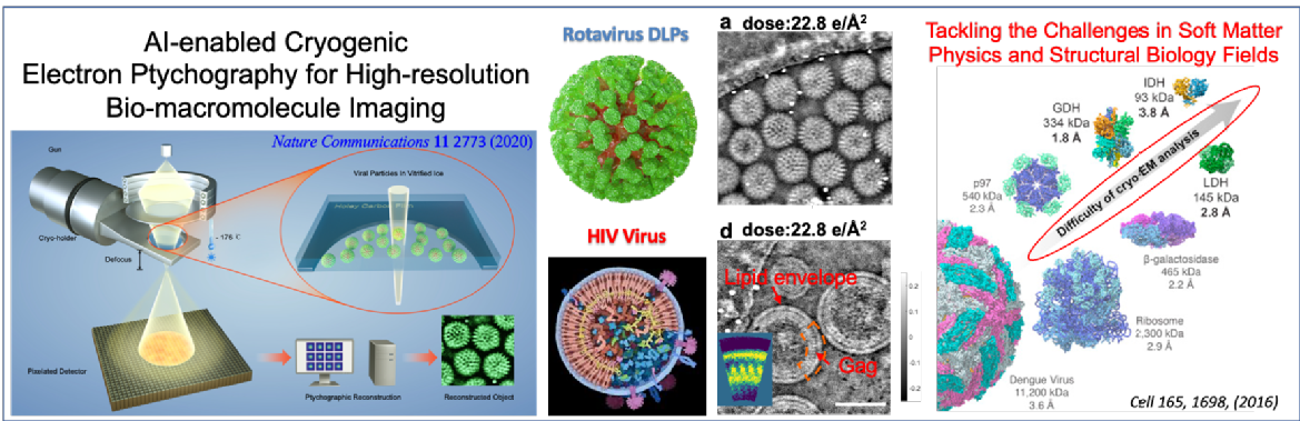
Cryogenic transmission electron microscopy (cryo-EM) with single-particle analysis (SPA) is a powerful method for visualizing a wide range of biological macromolecules in three dimensions (3D) at near-atomic resolution, which can provide direct insights into function and mechanism. Despite these revolutionary advances, it remains a challenge to deal with such small, heterogeneous and/or flexible molecules or complexes. An emerging strategy is based on cryogenic electron ptychography, which has been recently demonstrated by us for phase reconstruction of biological samples under low dose conditions. Ptychography is an emerging computational microscopy technique for acquiring images with resolution beyond the limits imposed by lenses, which has been applied to high resolution x-ray imaging. Due to its high phase-sensitivity, robustness to low electron dose data and the recovery of the sample wavefunction, electron ptychography represents a potentially disruptive change in the rapidly growing field of cryo-EM. The aim of this project is to provide a new computational image restoration framework for visualizing bio-macromolecules in 3D at near-atomic resolution. You will base upon artificial intelligence (AI) and machine learning techniques and develop a completely new computational scheme to dramatically speed up the time-consuming ptychographic reconstructions from a large quantity of electron diffraction, and recover high-fidelity phase-contrast images of biological macromolecules at high resolution. Combining with SPA, this AI-enhanced ptychography would potentially reveal and examine the 3D structures of the small molecules in an unbiased and comprehensive manner. You will be exposed to diverse and interdisciplinary research areas in applied mathematics, biological macromolecules, electron microscopy and cryogenics. You will experience an internationally collaborative environment, where you will closely collaborate with international-leading electron microscopists in the Rosalind Franklin Institute, a national research institute, dedicated to developing new technologies to tackle important health research challenges. You will have access to the state-of-the-art JEOL GrandARM with aberration correction, cryo stage, which is the very first and unique purpose-built cryo-EM in the UK for this category of work. You will also work with world-leading structural biologists in the Division of Structural Biology at the University of Oxford.