
Project Highlights
Nature Communications 14, 3027, (2023).
Cryogenic Electron Microcopy 3D Imaging
Many Congratulations to Xudong Pei, and coauthors! We would like to thank tremendous contributions from our collaborators to this work. The paper of Cryogenic electron ptychographic single particle analysis with wide bandwidth information transfer Xudong Pei1#, Liqi Zhou1,2#, Chen Huang3#, Mark Boyce4, Judy S. Kim3,5, Emanuela Liberti3, Yiming Hu1, Takeo Sasaki6, Peter D. Nellist5, Peijun Zhang4,7,, David I. Stuart4,7,Angus I. Kirkland3,5,7* and Peng Wang1,2* has been accepted by Nature Communications, 2023 May and is on line now.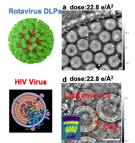
Nature Communications 8, 163 (2017)
Cryogenic Electron Ptychography
L Zhou (PhD student), et al, Low-dose phase retrieval of biological specimens using cryo-electron ptychography, Nature Communications, 11, 2773 (2020).
The need to determine the three-dimensional (3D) structures of biological macromolecules and assemblies at high resolution in their native states has motivated significant and sustained efforts to develop electron microscopy (EM) techniques, most notably phase contrast cryo-EM. However, unstained biological samples embedded in thin vitreous ice are essentially pure phase objects that are extremely radiation sensitive and consequently images of these have low signal-to-noise ratios and low contrast. To counter the latter, high defocus values can be used but these corrupt information transfer in single images at intermediate and high spatial frequencies due to rapid oscillations in the phase contrast transfer function.
In this project, we will develop an alternative method (Ptychography) based on scanning diffraction microscopy. This new approach provides tunable, continuous wide-band information transfer including low spatial frequencies that are inaccessible using conventional phase contrast imaging. Efficient phase recovery using ptychography provides higher signal-to-noise data than phase contrast imaging in cryo-TEM, which potentially reduces the particle numbers required for 3D reconstruction, facillitating 3D classification of heterogeneous specimens at low concentration or from structures with low symmetry.
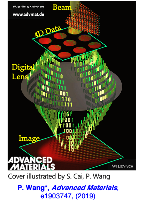
Data-driven Next-generation Electron Microscopy
Ptychographic diffractive imaging is one of coherent diffractive imaging (CDI) techniques to recover the object from diffraction data using a numerical solution to the phase problem. It uses a localized illumination probe moving across a sample while multiple diffraction patterns are recorded from overlapping areas as shown schematically in Fig. 1a. This data acquisition records redundant information and thus overcomes many of the limitations of conventional CDI reconstructions including non-unique solutions and a limited field of view. This data acquisition geometry also enables successful reconstruction without the requirement for a priori information about the object. Recovery of local phase changes at atomic resolution can provide structural data directly related to the properties of many important materials and a suitably sensitive, quantitative phase map can assist structural characterization at the atomic scale. Now, 2D Ptychographical Imaging at atomic resolution can be achieved and now it has been extended to 3D.
Physical Review Letters,121, 146101 (2018)]
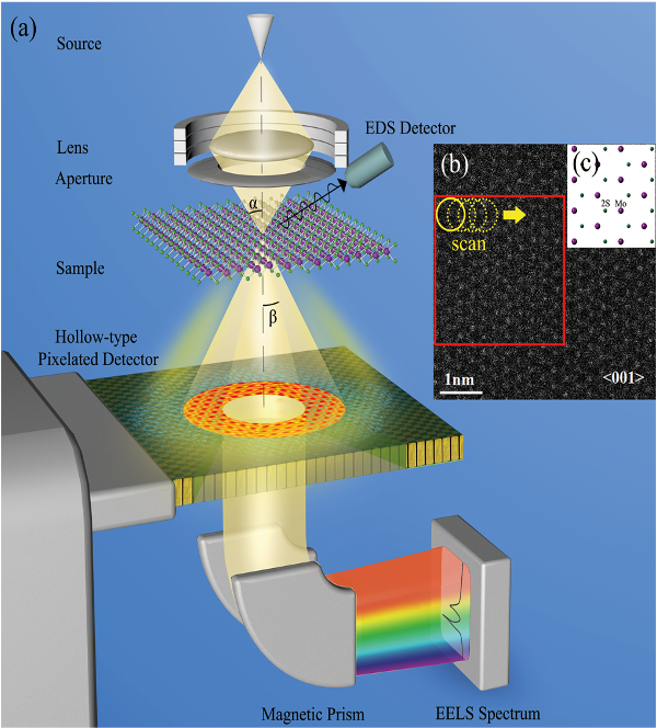
Multi-channel 5D STEM
B Song (Msci student), et al, Hollow Electron Ptychographic Diffractive Imaging, Physical Review Letters, 121, 146101 (2018).
Conventionally, TEM phase imaging which is sensitive to light atoms cannot be coupled with electron energy loss spectroscopy (EELS). With a fast hollow-type detector, we have conceived and designed a novel STEM imaging system based upon ptychography that can enable simultaneous, correlative analysis using both highly sensitive phase information and EELS chemical mapping at the atomic resolution. This work has been positively cited in literatures. For example, “Song et al. (2018) showed that even with part of the measurement signal removed, atomic-resolution phase signals can still be reconstructed. This “hollow” detector configuration is compatible with a large number of 4D-STEM techniques discussed in this paper and we expect it to find widespread use once dedicated “hollow” pixelated STEM detectors are widely available.” quoted from a Review article “Four-Dimensional Scanning Transmission Electron Microscopy (4D-STEM): From Scanning Nanodiffraction to Ptychography and Beyond” published in Microscopy and Microanalysis, 25, 563 (2019).
Nature Communications 8, 163 (2017)
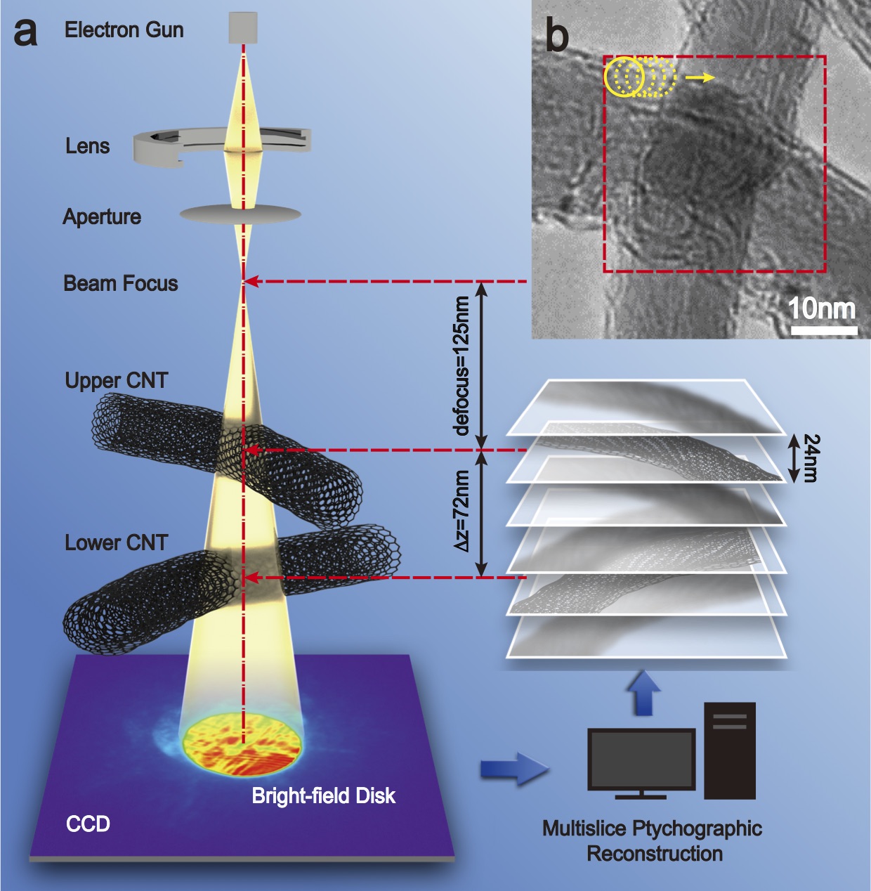
3D Multislice Electron Ptychography
S Gao (PhD student), et al, Electron ptychographic microscopy for three-dimensional imaging, Nature Communications, 8, 163 (2017).
Ptychography originally was based on a 2D multiplicative approximation to model the interaction of the probe and the specimen. However, this approximation breaks down at modest specimen thickness for electrons due to their larger interaction cross section, which leads to significant multiple scattering. In this work, we implemented a multislice method to correct for multiple scattering in 3D phase reconstructions by treating the 3D object as a series of 2D slices, each with its own transmittance function, separated by small distances of free-space propagation. With this strategy, we were able to demonstrate for the first time that defocused probe electron ptychography can provide high contrast, quantitative phase information from a 3D structure of a thick sample (including amplitude and phase) at close to atomic lateral resolution and with a few tens of nanometers depth resolution. This 3D reconstruction approach is potentially applicable to in situ TEM experiments as it does not require mechanical tilting of the specimen through large angles as is the case for electron tomography.