Imaging Facilities

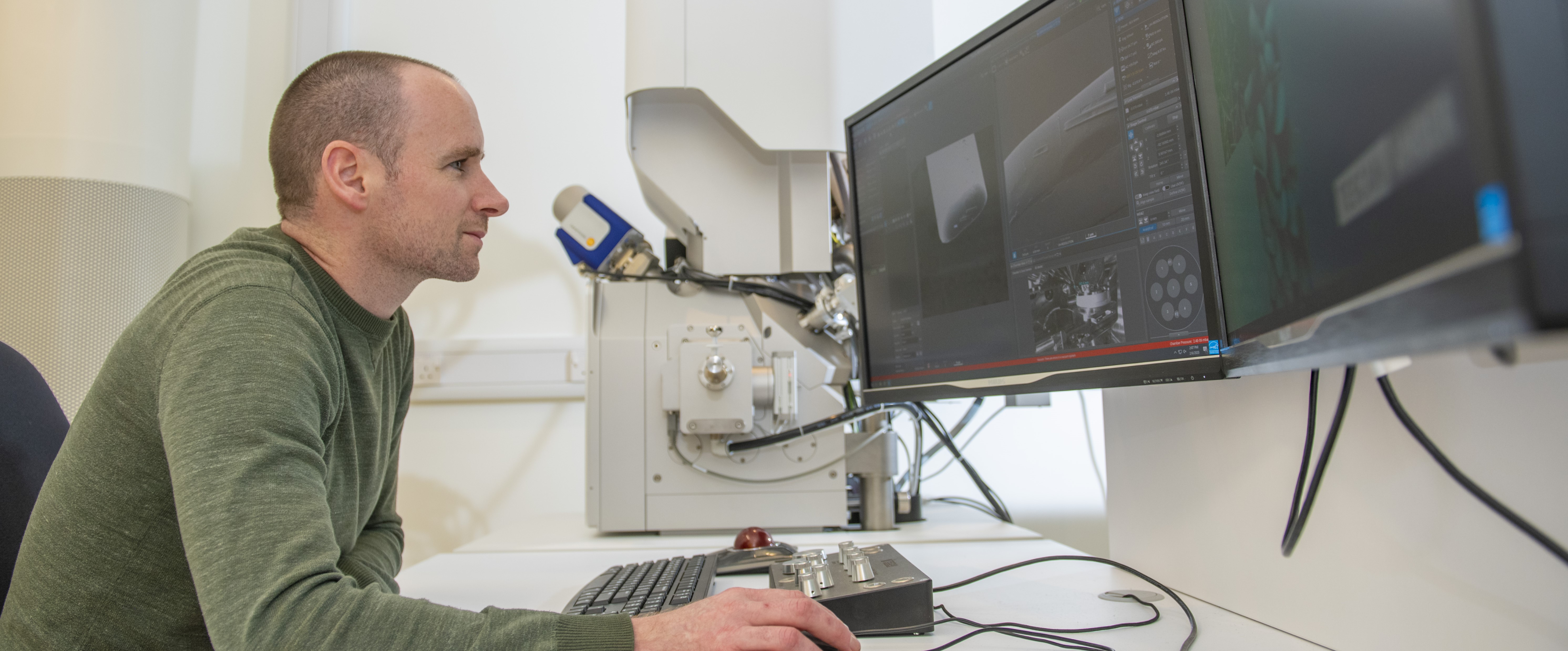
Whether you are looking for in-depth information about the structure or surface of a material or ultrastructure imaging of biological samples, we have a wide selection of imaging services available to suit your needs. Three of our specialist Research Technology Platforms (RTPs) are dedicated imaging facilities: the Electron Microscopy RTP, the X-ray Computed Tomography RTP and the Advanced Bioimaging RTP.
In addition, the Computing and Advanced Microscopy Development Unit (CAMDU)Link opens in a new window at Warwick Medical School houses cutting-edge imaging systems and powerful analytical resources that are driving innovation in biomedical and medical science.
Please contact Claire Gerard () to discuss your requirements.
Scanning Electron Microscopy
Our Electron Microscopy Facility houses three Scanning Electron Microscopes that can image surfaces with a wide range of magnifications and resolutions at nanometre scale.
Transmission Electron Microscopy
We have three TEMs capable of up to atomic-resolution imaging. They transmit electrons through an ultra-thin specimen to form an image of its structure. Scanning Transmission Electron Microscopy (STEM) is also available.
Atomic Force Microscopy
The Atomic Force Microscopes in our Electron Microscopy facility can be used to obtain high-resolution images showing topography, surface adhesion and electronic properties of a wide variety of sample types.
Focussed Ion Beam Lithography
Cross-sectioning and TEM sample preparation can be performed using our focused ion beam scanning electron microscope.
X-ray Computed Tomography
Our XCT imaging facility creates 3D volumetric models of samples, with machines ranging from high penetration for large metallic objects to an ultra-high resolution system for imaging nanometre features.
Computing and Advanced Microscopy Development Unit
The Computing and Advanced Microscopy Development Unit (CAMDU) at Warwick Medical School houses cutting-edge imaging systems and offers dedicated image analysis services.
Advanced Bioimaging
The Advanced Bioimaging Facility supports the investigation of complex biological problems using electron microscopy. It specialises in transmission electron microscopy (TEM) including Cryo-TEM, thin-section TEM and negative-stain TEM.
WMG Microscopy Suite
The microscopy suite in Warwick Manufacturing Group houses a wide range of microscopes, including SEM, TEM and AFM capabilities, as well as a range of sample preparation equipment. The lab is particularly well equipped to support steels research and battery research.
Confocal Microscopy
Confocal microscopes use a pinhole to collect light, discarding out-of-focus light to produce clear, high resolution images. Confocal microscopy is widely used for analysis across biological disciplines.
Optical Microscopy
We have a wide selection of optical microscopes for work with cells, tissues and material samples. We offer fluorescence microscopy for illuminating live cells and have a 3D optical microscope in our metrology lab.
Catholuminescence
A cathodoluminescence system is attached to a scanning electron microscope (SEM) and can be used for minerals analysis, investigating chemical composition and revealing stress information in semiconductors and insulators.
