WMS Events Calendar
Please see this page for MB ChB events.
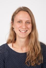
BMS Seminar: Building and breaking epithelial tubes, Dr Clare Buckley, Department of Physiology, Development and Neuroscience, University of Cambridge
Abstract: My group strives to understand how mechanics and biochemical signalling work together to shape epithelial organs during development. We are currently particularly interested in understanding the links between apical-basal cell polarity, cell-cell adhesion, and cellular mechanics during the process of vertebrate secondary neurulation and mammalian early epiblast formation. Both develop from a relatively simple epithelial tube/cavity, containing a central, fluid-filled lumen. However, rather than adopting the well characterised folding mechanism of lumenogenesis to ‘close’ an already epithelial structure, both structures initiate via de novo apical-basal polarisation (mesenchymal to epithelial transition, MET) within the centre of an initially solid organ primordium, followed by hollowing (opening) of the epithelium. To study these processes, we use high resolution imaging and optogenetic approaches in vivo, within the developing zebrafish neural tube and in vitro, within multicellular mouse embryonic stem cell (mESC) cultures. This allows us to quantitatively image the behaviour of cells before, during and after a precise manipulation in, for example, actomyosin contractility at a subcellular scale. We have recently found a new Cadherin-mediated mechanism of apical membrane initiation site (AMIS) localisation within de novo polarising epithelial tubes/ cavities. This suggests that cell-cell adhesion acts as the first symmetry-breaking event during tube organogenesis.
 Biography: Clare’s undergraduate degree was in Biological Sciences at Oxford. During her PhD she developed models of myelination in zebrafish with Robin Franklin and DanioLabs at Cambridge. She became obsessed with apicobasal polarity and epithelial tubes during her postdoc with Jon Clarke at KCL and hooked on optogenetics during a visiting scholarship to Orion Weiner’s laboratory at UCSF. She started her lab at Cambridge with a RS Dorothy Hodgkin Research Fellowship and is now a WT/RS Sir Henry Dale Fellow. Her lab uses optogenetics and live confocal imaging to investigate the morphogenesis of epithelial organs during development and disease.
