
Graphene membranes for microscopy
 |
Graphene is widely known as a modern wonder material. Despite being only a single atom thick, it has amazing electrical properties and exceptional mechanical strength. This has led to many predictions of possible future applications, but there is an area of science in which it is already making a significant practical difference. |
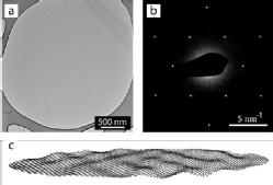
The atomic structure of materials defines their physical properties. This is true as much for understanding and developing new nanoparticle catalysts as it is for understanding the function of proteins and enzymes. With significant recent advances in microscope hardware, our ability to investigate this nanoscale structure has been transformed over the last 5 to 10 years - there has been a resolution revolution. The standard approach is to use transmission electron microscopy, where a beam of electrons is passed through the material (nanoparticle, molecular assembly etc.) and the interactions of the electrons with matter are used to discern the structure. To do this, the material must be placed on a membrane and the structure of the membrane contributes to, or rather detracts from, the measurement. Often, it is this support membrane that limits the resolution that can be attained. At a single atom thick, graphene is the perfect material for this support membrane. At Warwick, we were among the first to develop the use of graphene membranes for microscopy and we have worked with companies such as EMResolutions to develop a commercially viable method for their production. These single atom thick membranes are now used worldwide to help solve scientific problems in areas such as medical research and medical testing, development of renewable energy and battery technology, and green chemistry approaches to chemical synthesis.
For more information, contact Dr Neil Wilson.
Links to some of our underpinning research
'Graphene Oxide: Structural Analysis and Application as a Highly Transparent Support for Electron Microscopy.'
N.R. Wilson, P.A. Pandey, R. Beanland, R.J. Young, I.A. Kinloch, L. Gong, Z. Liu, K. Suenaga, J.P. Rourke, S.J. York, and J. Sloan,
ACS Nano 3 (9), 2547 (2009).
'Imaging the Structure, Symmetry, and Surface-Inhibited Rotation of Polyoxometalate Ions on Graphene Oxide.'
J. Sloan, Z. Liu, K. Suenaga, N.R. Wilson, P.A. Pandey, L.M. Perkins, J.P. Rourke, and I.J. Shannon,
Nano Letters 10 (11), 4600 (2010).
'A simple approach to characterizing block copolymer assemblies: graphene oxide supports for high contrast multi-technique imaging'
JP Patterson, AM Sanchez, N Petzetakis, TP Smart, TH Epps III, I Portman, ... and NR Wilson
Soft Matter 8 (12), 3322-3328 2012