
Claire Fletcher
I am studying for a PhD part time while working in the Radiotherapy department for University Hospitals Coventry and Warwickshire NHS trust (UHCW).
PHD Project Title: Development of cone beam computed tomographic biomarkers for lung radiotherapy
Project Supervisors: Dr Jon Duffy and Dr Jane Rogers
Project Summary:
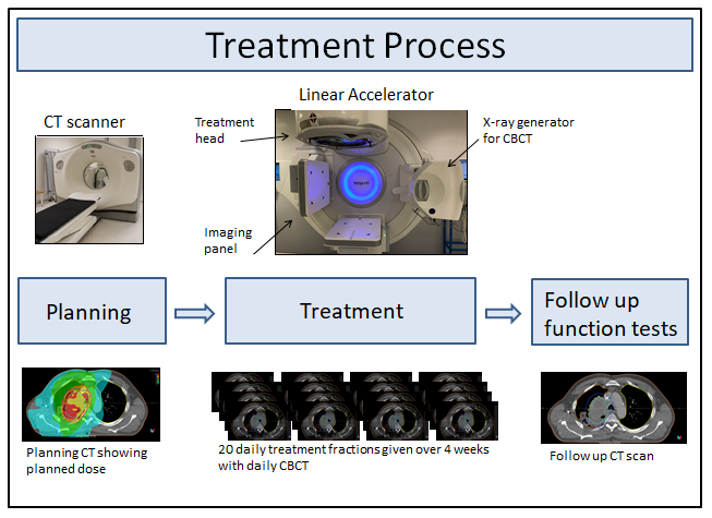
Radical radiotherapy is commonly used for inoperable, non-metastatic lung or oesophageal cancers, and radiation-induced lung damage is recognised as a complication. The radiotherapy treatment usually given in the UK consists of 55 Gy in 20 fractions which are delivered to the patient once a day over 4 weeks. A cone beam CT (CBCT) is performed on each visit immediately prior to each treatment fraction in order to position the patient accurately. During the course of treatment, a variety of changes are frequently seen in the either the tumour or lung. Although these CBCT images are currently only used for positioning, there is potential in using the images to adapt the ongoing treatment strategy, which is not something which is currently common practice.
This project will use CBCT data of patients treated at University hospitals Coventry to correlate patient outcomes with changes in CBCT images during the treatment course. The patient data being used to develop the methodology will be from patients who have already completed their treatment. My research project will involve developing the analysis technique. The changes to both the tumour and normal lung tissue will be analyzed using a variety of techniques in order explore correlations in changes seen during treatment and patient outcomes.
The aim of the project is to determine parameters which can be further investigated clinically to potentially adapt radiotherapy during the course of patient treatments to reduce toxicities and/or increase tumour control.
Radiotherapy Treatment Process:
Current Progress:
- CBCTs from anonymised historic patients were analysed to develop and test research methodologies.
- CBCTs were corrected for daily fluctuations in image intensity
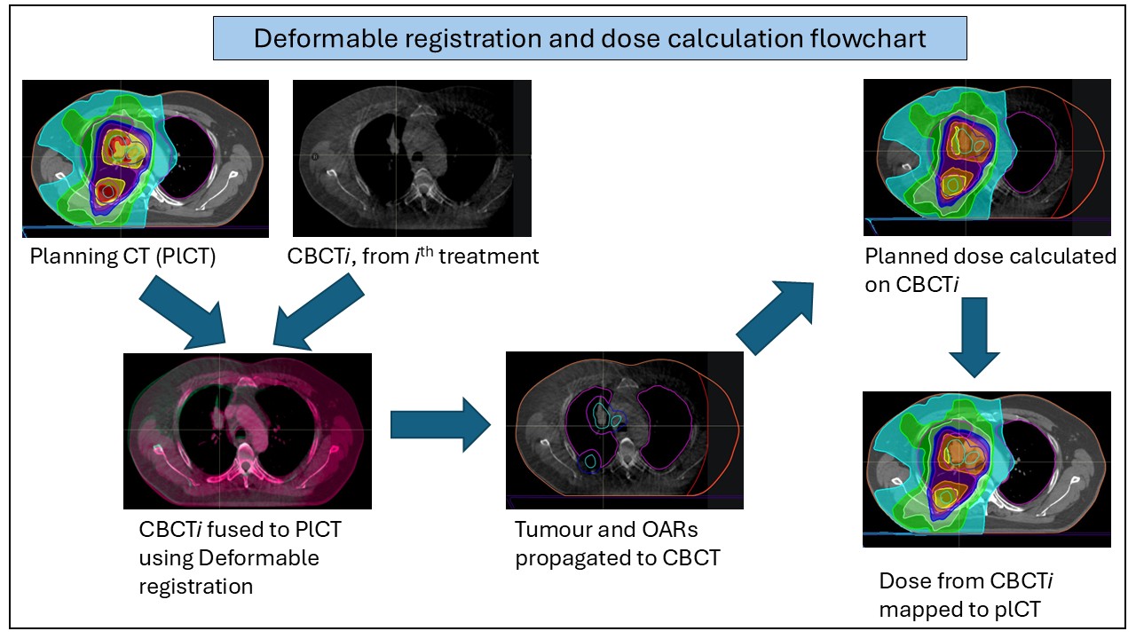
- Deformable registrations using RayStation were used to register CBCTs to planning CTs.
- Python scripts were developed to automate the registration of images and propagation of the defined lung and tumour volumes
- These corrected CBCT images were used to analyse lung density and tumour volume changes.
- Planned doses were recalculated on CBCTs and the dose distribution deformed in order to assess treatment doses received by healthy lung.
- Metrics such as the volume receiving 5Gy and 20Gy dose to the lung were calculated. As well as the mean lung dose and the equivalent uniform dose to the lung.
- The point during treatment at which these metrics could be reliably used to predict the final values was determined.
- The effect of adapting treatment at different points during treatment was determined in order to find the optimal adaptation point.
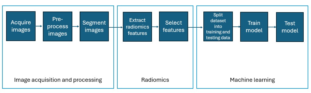
- Radiomics Features were extracted from CBCTs in order to look at feature changes during the course of treatment
Future Work
- Investigate correlations between metrics from CBCTs to lung function tests and the risks of lung damage.
- Use Machine learning models to evaluate radiomics features and their use in predicting response to treatment.
Glossary
Radiotherapy: The destruction of cancerous cells with high energy (megavoltage, MV) x-rays
Fractionation: Radiotherapy treatments are delivered in a number of small fractions, usually one a day, in order to allow normal healthy tissues to recover while maximising destruction of cancerous cells
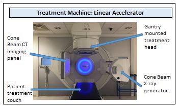
Linear Accelerator: Machine used to deliver radiotherapy using a gantry moving around the patient.
Planning CT (PlCT): Before treatment by radiotherapy a patient will be given a planning CT scan in order that the treatment can be planned using computer software (a planning system)
Adaptive Radiotherapy: Patients can be re-planned based on changes occurring over treatment, for example tumour shrinkage
Cone Beam CT (CBCT): CBCT uses a cone shaped kilovoltage (kV) x-ray beam and 2D detectors (instead of the usual fan shaped x-ray beam and 1D detectors used in conventional CT scans), this allows volume images to be acquired with just one rotation of the x-ray gantry
Deformable Image Registration: Comparison between two images, where the target image is warped to match the reference image
Radiomics: Data extraction from medical radiological images which is undetectable by the naked eye and describes the images in terms of minable data including histogram and intensity information as well as data relating different voxels to each other