
Positions
A PhD project (funded, Start date: October 2023) is available
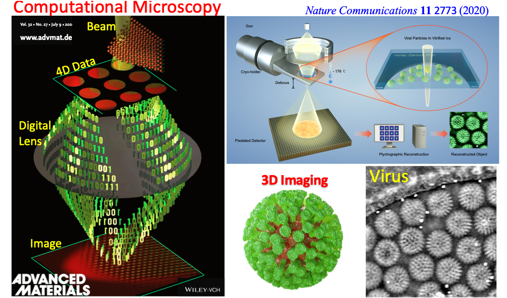
Low-dose phase retrieval of biological specimens using cryo-electron ptychography. Nature Communications 11, 2773, (2020).
Cryogenic Electron Ptychography For Biological Imaging
The Nobel Prize-winning technique, cryogenic electron microscopy (cryo-EM) is a powerful method for visualising a wide range of biological macromolecules in three dimensions at near-atomic resolution, which can provide direct insights into function and mechanism. Despite these revolutionary advances, cryo-EM has been primarily based on conventional phase contrast TEM imaging at high defocus and suffers from heavy background noise and low contrast due to a limited electron dose used in imaging for reducing radiation damage to biomolecules.
Here, you will be developing a completely new computational microscopy so called cryogenic electron ptychography, further enhanced by artificial intelligence (AI) and machine learning techniques to recover high fidelity amplitude and phase contrast images of biological macromolecules with state-of-the-art ultrafast detectors at low doses.
In this project, you will have strong collaborations with the scientists in the Rosalind Franklin Institute, dedicated to developing new technologies to tackle important health research challenges and the structural biology experts in the Division of Structural Biology, University of Oxford.
The application deadline for all courses starting in September/October is 23:59pm (GMT) on 20 January 2023 however; places are limited, so you should submit your application as soon as possible.
A PhD project (funded, Start date: October 2022) is closed
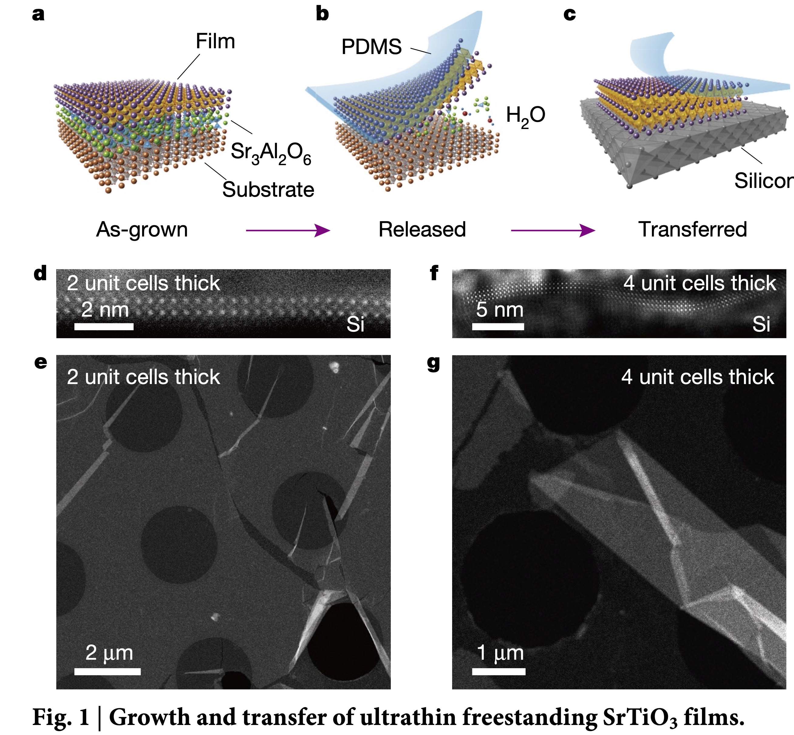
Freestanding crystalline oxide perovskites down to the monolayer limit. Nature 570, 87-90, (2019).
High-Resolution Probing Ferroic-ordering by Cryogenic Electron Ptychography
The emergence of ferroic orderings in materials at the atomically thin limit, including ferromagnetism and ferroelectricity, has attracted tremendous attention due to their novel physics and promising applications for future flexible nanoelectronics including artificial e-skin, flexible touch sensors and health monitors. Understanding the underlying physics behind the ferroic phenomena requires a high-resolution image technique that can directly visualize magnetic or electric fields in the materials at the nanometer, even down to atomic resolution.
The aim of this project is to develop a new pioneering algorithm-driven imaging technique called ptychography in conjunction with machine learning and ultrafast detectors. Much of the work will involve using scientific Python or Matlab to develop new ways of quantitatively analysing ferroic properties and structure in 2D ferroic thin films at the atomic scale. Combined with tomographic methods, we will further extend 2D field projections into 3D vector field reconstruction. Furthermore, we will look at dynamic behaviours of materials in situ and study how they respond to a changing external stimulus (electric, magnetic field, temperature).
The experimental part of the project will be based in the Warwick Analytical Science Centre, which hosts a state-of-the-art JEOL ARM microscope where a new cutting-edge ultrafast detector allowing the image to be recorded at 1000 frames per second can be applied in a very wide range of 4D STEM image techniques such as virtual STEM, DPC etc..
The application deadline for all courses starting in September/October is 23:59pm (GMT) on 20 January 2022 however places are limited so you should submit your application as soon as possible.