Skip to main content Skip to navigation

Homepage contents

Course Content Page

3D printed models to aid the jury

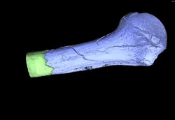
We received a section of an individual’s skull which showed the impressions of some form of instrument. The micro-CT displayed these impressions in great detail, revealing two distinct shapes. These could be matched with a hammer and a spanner that were found at the scene, proving that both tools had been used.

The skull section was 3D printed and shown to the jury during trial to visualise the injuries. This was accompanied by Professor Williams giving expert witness testimony regarding the scanning and 3D printing processes to validate the evidence.

3D scanning and imaging supporting evidence of child abuse

This was a complex case of suspected child abuse. Multiple parts of an 18-months-old infant were scanned at WMG following the detection of several injuries at the hospital. The right femur (thigh bone) showed a grossly displaced fracture with a large amount of callus forming around it, indicating that healing processes had begun without proper medical attention. The ribcage displayed twenty individual fractures in different stages of healing. Images of these fractures were also shown at court where it has been established that the child’s guardian had caused the injuries in at least three separate events.


Establishing cause of death

In the case of the unexplained death of a young woman, we provided 3D scanning evidence to assist the pathological diagnosis of cause of death. The technology revealed damage to the victim's larynx, in detail far beyond the capabilities of medical CT scans, that confirmed that the victim had been forcibly strangled. This evidence contributed to the conviction of the offender for murder.

Homicide - complex dismemberment

The two pieces of humerus fit togetherIn a case of complex dismemberment, we delivered one of the first examples of micro-CT technology as a forensic radiological method in a UK courtroom.

Asked to examine a charred piece of evidence thought to contain human bone, we discovered that it was a perfect jigsaw fit to another piece of bone in the case, and were able to show the tool marks on both pieces in micro scale (one 50th of a millimetre). These matched the characteristics expected for the type of saw the offender had disposed of alongside the victim.

Our 3D printed models of the two pieces were able to demonstrate the evidence to the jury.

Identifying weapon from skeletal remains

A request was made to WMG to scan several bones of a skeleton found during construction works. There were signs of dismemberment on the bones and the affected areas were therefore micro-CT scanned to gain information regarding the weapon used.

The scans depicted subtle V-shape grooves around the main joints and the neck. This indicated two things: that a knife had been used and that the perpetrator must have had some knowledge of anatomy or butchery.

Infant skull-fracture shown to be the result of a single impact

In this case, we scanned an infant’s skull bones which displayed a prominent fracture attributed to non-accidental circumstances. Investigators sought to verify if further cracks on the skull were actual fractures or additional sutures which would represent normal anatomical features.

The scan images clearly showed that the latter was the case, indicating that the injuries were the result of a single impact and not multiple.

Model for facial reconstruction

We were asked to scan a skeletonised skull belonging to an unidentified individual found along the M54. Police hoped to use the resulting model as the base for facial reconstruction to help identify the deceased. The model showed a healed nasal fracture and some dental restorations. Ultimately, DNA was used to identify the individual and the facial reconstruction was not completed.

Road Traffic Accident

RTA 3D model
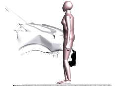
In the case of a fatal Road Traffic Accident, 3D imaging technology allowed us to precisely recreate multiple elements of the crime scene, and put together a complete picture of what took place.
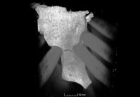
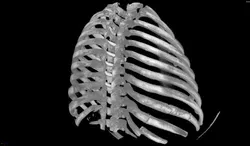
We provided vital evidence explaining an injury to the victim that was not typically consistent with an RTA. Using novel techniques, a superficial scratch mark on the victim's leg bone was identified, which had been missed on the hospital CT, and which was matched with a spike on the front wing of the vehicle. Due to our evidence, it was possible to conclude that the victim was impaled on the front of the vehicle at the point of impact.

Our virtual model of the whole crash scene, achieved by laser scanning at different scales, was presented in court.

Ruling out foul play

A postmortem examination revealed two fractures at the base of a 64-year-old's skull only centimetres apart, which did not appear consistent with a fall. At the scene, a damaged front door and signs of a struggle in the hallway meant detectives suspected foul play.

A precise 3D printed model of the skull was produced in our lab and taken to the scene, where it showed perfect matches with the geometry of the door handle. This suggested that the individual had fallen, twice hitting his head. The model therefore aided establishing an accidental cause of death.

Ruling out violence as the cause of a suspicious death

We scanned the neck structures of a male individual whose death appeared suspicious despite the absence of any evidence of violence. Subtle changes raised the possibility of compression of the neck but the larynx was found to be intact on the micro-CT scans. Third-party involvement was therefore excluded and no criminal investigation ensued. The cause of death was later confirmed as drug-related.

Video animation of knife injury to illustrate evidence

Knife woundPolice sought WMG’s assistance to scan a sternum (breast bone) containing a stab wound. They wanted to determine what type of knife had caused the injury.

The micro-CT scan showed a clear outline of the weapon which had penetrated the entire thickness of the bone. A video animation of the injury was shown at court to illustrate this

Visualisations to assist in specialist testimony

Ribcage - healed injuriesIn a case of suspicious infant death, our scans revealed, in microscopic detail, that the victim had suffered a series of injuries which showed various degrees of healing without signs of medical intervention. This indicated neglect and abuse over a period of time.

The 3D visualisations of the injuries were used to illustrate the pathology expert’s testimony in court.

The child’s legal guardian was found guilty of murder and sentenced for life.

  Understanding the circumstances of an assault

We scanned the neck structures of a woman who was found deceased in her bath. Witnesses reported hearing an argument between her and her estranged partner. The forensic post-mortem revealed multiple injuries, making a diagnosis rather complex. Subtle fractures seen on the micro-CT images added that compression of the neck would have contributed to the event. Whilst not the ultimate cause of death, it improved the understanding of the circumstances of the assault.