Linear Dichroism
LD is a spectroscopic technique primarily used to probe secondary structure of proteins and DNA.
Measurement of LD
To measure an LD spectrum the sample requires orientation. This is usually achieved using shear flow, linear flow or a stretched film.
An LD spectrum is found by taking the difference between absorbance parallel and perpendicular to the axis of orientation, so
![]()
Applications
LD can be used to measure absorbance spectra or kinetics. Below is a typical DNA spectrum for LD. The minimum at 260 nm is from pi–pi* transitions along the DNA backbone.
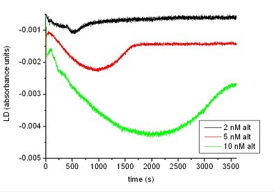
We have also measured kinetics. Below is a measurement of the restriction enzyme BstZ17I acting on different concentrations of a pCDNA3.1 derivative plasmid.

Linear dichroism is also applicable to DNA groove binding molecules, fibrous proteins and membrane proteins.

A linear dichroism flow cell. The sample is placed in a quartz microcuvette. A rod is inserted and the cuvette rotated which aligns the sample by creating a Couette flow.
