Visitor Programme: Warwick's Lattice LightSheet Microscope
The Wellcome funded Lattice LightSheet Microscope is available for for visitors to carry out their own experiments.
To register an initial interest please fill out this form. We will then contact you to discuss the details of the project and arrange time on the microscope. Feel free to email David Corcoran, the Lattice Lightsheet Specialist, to ask questions (david.corcoran@warwick.ac.uk).
Technical specifications and sample requirements can be found on the instrument page.
Separately from the Visitor Programme, collaborators of CMCB researchers are welcome to use the lattice for joint projects. You will be hosted by your collaborator who will facilitate the experiments on the lattice. Please contact CAMDU for more details (camdu@warwick.ac.uk).
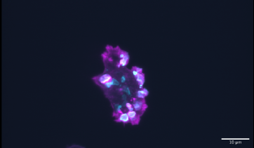
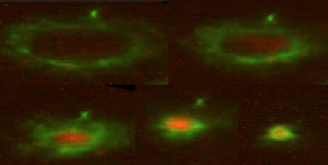
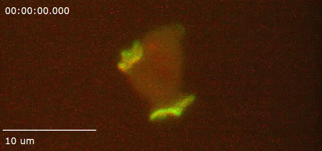
Recent Visitors
|
Dr Rob Kay |
The work on macropinocytosis we are doing with Helena Coker at CAMDU is the focus of my current research. Macropinocytosis – an ancient and conserved way for cells to take up large volumes of medium into internal vesicles – is used by cancer cells and amoebae for feeding. We obtain spectacular movies of macropinocytosis in Dictyostelium by LLSM, which can be obtained in no other way. This allows us to see how macropinocytic cups form and close. From small beginnings, we are now mapping a significant number of reporters through the life of a cup and in a number of mutants defective in the process. This has become our Macropinocytosis Mapping Project. Thanks Helena! |
|
Dr Paolo D'Avino |
We used the Lattice Light Sheet high resolution microscope at the Computing and Advanced Microscopy Unit (CAMDU) of the Warwick Medical School to visualise by multidimensional time-lapse imaging the dynamics of two fluorescent-tagged proteins, Citron kinase and the kinesin KIF23/MKLP1, during cytokinesis in human HeLa cells. We were very impressed by the high quality and resolution of the images as well as by the very low photo bleaching. Our entire experience with CAMDU was excellent and we were particularly impressed by the technical ability, availability and professionalism of the Imaging Specialist, Helena Coker. It just thanks to her that we were able to obtain such stunning time-lapse images. We are planning to go back to CAMDU as soon as possible and it is our hope that the CAMDU and their visitor programme will continue to receive strong support from the Wellcome Trust. |
|
Professor Igor Weber |
I spent 5 days using the Warwick Lattice Light Sheet microscope for studying spatio-temporal correlation between activated Ras and its negative regulator IqgC in live Dictyostelium cells. Due to unique capability of the LLS microscope to acquire fully resolved cell volumes in two fluorescence channels every 2 seconds. We unraveled several features of intracellular protein dynamics that were not apparent using conventional confocal microscopy. |
|
Dr Jason King |
Being able to come to Warwick and use the Lattice light sheet has been fantastic, enabling us to observe dynamic cellular events in our highly photo-sensitive cells in far more detail than we have been able to before. The set up in Warwick has been superb and in particular, the skill and experience of their imaging specialist Helena has been invaluable to enable us to make the most of the system and obtain excellent data and beautiful movies right from the start. This has really expanded our biological understanding, and will hopefully be the basis of many future projects. |