
Slime moulds under the microscope
Slime moulds aren't just bags of goo, inside they have some complex anatomy. Just like our bodies have different parts that do different jobs like lungs, spleens and eye, cells have different parts as well - in a body we call them organs, in cell they're little organs - organelles. Slime moulds may be weird but they have the same basic parts as other cells.
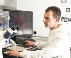
Confocal Microscopes
Confocal Microscopes scan a laser over the sample but we don't look at the laser light reflected back. Scientists label antibodies and molecules with fluorescent dyes that only stick to the parts of the cell we want to look at. The laser makes the dyes fluoresce like a highlighter pen and the microscope only sees the fluorescent signal so we can highlight only the bits of the cell we're interested in.
The Cytoskeleton
All cells, not just slime moulds have an internal scaffolding called the cytoskeleton, it's a network of protein fibres that control the cell's shape. In a slime mould - which is always crawling around looking for food, the cytoskeleton constantly rearranges - fibres get broken down in at some spots and rebuilt at others pushing the cell this way and that.
The poison phalloidin from death cap mushroomsLink opens in a new window sticks to the cytoskeleton and stops it moving, in the lab we stick a fluorescent molecule to phalloidin and use it to highlight the cytoskeleton.
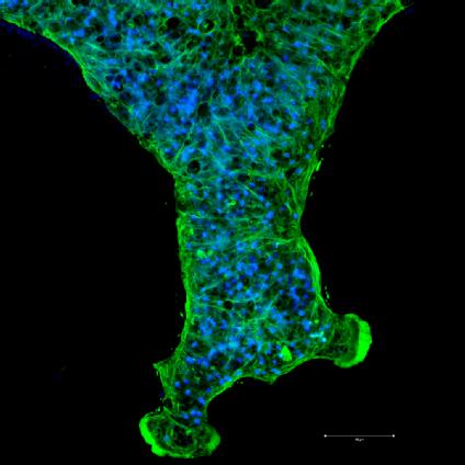
The blue are the nuclei which contain the cell's DNA. You can see there are thousands of them in this one tiny piece - a normal cell has one nucleus, giant cells have to have thousands of nuclei to coordinate and control cell activity.
The green is the cytoskeleton - in most of the picture it's in long thin strands across the vein, these fibres expand and contract making the slime mould pulse and moving liquid around the cell. Towards the bottom middle and right where it's fanning out you can see much denser areas of solid green - these are areas where the slime is moving quickly and loads of cytoskeleton proteins are piled up ready to make new scaffolding for the cell.
The dark round holes in the green round holes are pores in the surface of the cell - some are to let slime exude out and leave a trail, some are there to let the slime sample the outside environment as it searches for food.
Live Imaging
To look at the cytoskeleton we have to 'fix' the cell - that's a fancy way of saying kill it very carefully - to see that fine detail we need everything to stay still so we treat the sample with formalin which kills the cell but also fixes all the proteins together so they make a snapshot of exactly what the cell was doing. With a light microscope we can look at live things though.
In this video - We've stained the cell membrane with a green dye. The mitochondria are stained red. The slime has been cut - yellow is where popped mitochondria and torn cell membrane get mixed up - but you can see it doesn't take long to put itself back together and reconnect veins.