
Electron Microscope
Light microscopes are great, they make beautiful pictures and allow us to look inside living things but there's a limit to how much they can magnify things. If you want to see stuff that's really small you need to use an electron microscope.
Electron microscopes can magnify things hundreds of thousands of times but they need something that's very thin so we can shine an electron beam through it, and very dry because they work in a vacuum and water would just boil away. We dry our slimes out in alcohol and set them in a resin that hardens when it's baked.


The resin embedded slimes are sliced on a machine called an Ultramictome which uses a diamond knife into cut pieces a hundred times thinner than a human hair ready for us to put in the microscope.
Overview
Cells are very complicated with many parts. With an electron microscope, even a single picture can contain a huge amount of information.
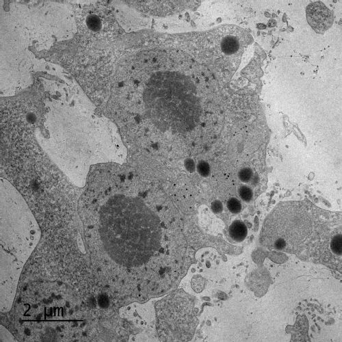
This is a slice through a slime mould vein - they're very busy inside. Electron microscopes only make black and white images but we often colour them in to make it easier to see what's going on. Have a look below to see a labelled version

Membranes - there are a lot of membranes in cells - anywhere you see a boundary with a thin dark line on this picture is a membrane. They're made of oils and keep different parts of a cell separated. If you look closely around the nucleus you can see it has a double layer and tiny gaps called nuclear pores to control what goes in and out of the nucleus.
Ectoplasm - ordinary cells just have 'cytoplasm' - the goo inside that contains everything. Slime moulds have two layers - the ectoplasm is the bit with all the normal cell stuff in it - it's on the outside layer of the veins.
Endoplasm - the other layer, it's much more liquid than the ectoplasm, it's the fluid in the veins and contains dissolved food and other goodies.
Endocytosis - cell membranes are constantly on the move. To get something big (like a tasty bit of oat flake) inside a cell they can fold in and form a funnel around the target that pinches off to make a tiny bubble called a vesicle. The opposite - spitting things out is called exocytosis.
Mitochondrion - These organelles make energy for the cell - they break down sugar and use it to make ATP which is a sort of portable energy source for chemical reactions in the cell.
Nucleus - the bit with the DNA, it takes in information from the rest of the cell and sends out instructions on what proteins to make. The dark patches are nucleoli - areas where the DNA has been opened up for copying.
Lysosomes - special vesicles that contain powerful digestive enzymes that digest and reuse old and damaged bits of the cell.