
Automatic Classification of Hyperspectral Images of Human Colon Biopsy Slides
Bowel cancer is the third most commonly diagnosed cancer in the UK after lung and breast cancer. It is the second most common cause of cancer death after lung cancer accounting for over ten percent of all cancer deaths. In the UK alone, there were over 35,000 colorectal cancer (a combined term for colon/rectum cancer) cases in the year 1999 and more than 16,000 deaths from bowel cancer in year 2000.
Our research is aimed at developing efficient algorithms for automatic analysis of microscopic level image data cubes of normal and malignant (adenocarcinoma) human colon tissue in order to assist the pathologists with the diagnosis of colon cancer. The challenge is to automatically classify between normal and malignant tissue sections with an acceptable accuracy. Our focus in this area is on spatial analysis (as opposed to spectral analysis) and the application of various machine learning methods on features extracted by the spatial analysis.
 |
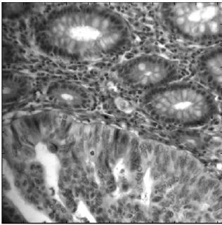 |
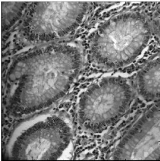
| Normal biopsy | Biopsy with a malignant gland |
Two sample human colon tissue images: The images were acquired by colleagues at Yale University (USA) School of Medicine from archival H & E (hematoxylin & eosin) stained micro-array tissue sections. The dimensions of each data cube were 1024×1024×20, where 20 spectral bands in the wavelength interval 450–640nm were used.
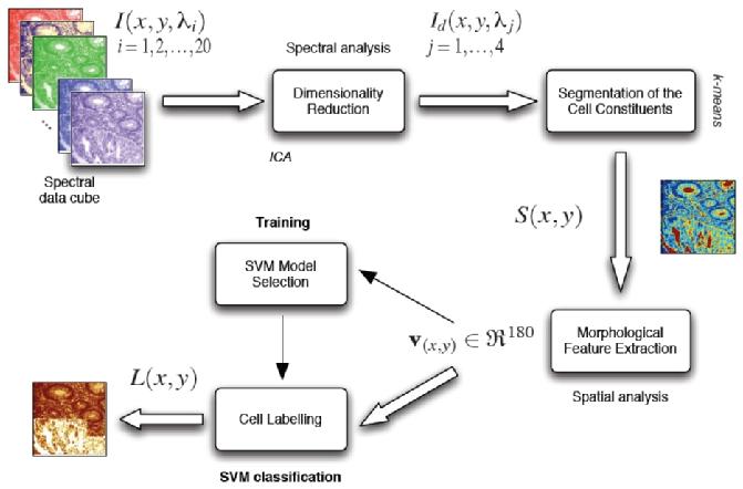
Kashif Rajpoot worked with morphological features on binary cell constintuent images obtained by segmentation using only 4 linearly independent component bands. An overall schematic diagram of our classification system, proposed initially in the MICCAI'2004 paper, is shown below.
Recently, Khalid Masood has been looking into various textural and shape-related features coupled with subspace projection methods for analysis of 128-band hyperspectral data cubes of colon biopsies acquired by the Yale colleagues using a novel DMD-based hyperspectral camera developed by Plain Sight Systems.
Relevant Publications
- K Masood, NM Rajpoot,
Classification of Colon Biopsy Samples by Spatial Analysis of a Single Spectral Band from its Hyperspectral Cube,
in Proceedings Medical Image Understanding and Analysis (MIUA'2007), July 2007

- K Masood, NM Rajpoot,
Hyperspectral Texture Analysis for Colon Tissue Biopsy Classification,
in Proceedings International Symposium on Health Informatics and Bioinformatics (HIBIT'2007), May 2007
- K Masood, NM Rajpoot, H Qureshi, KM Rajpoot,
Co-occurrence and Morphological Analysis for Colon Tissue Biopsy Classification,
in Proceedings 4th International Workshop on Frontiers of Information Technology (FIT'2006), 2006
- KM Rajpoot, NM Rajpoot,
SVM Optimization for Hyperspectral Colon Tissue Cell Classification,
in Proceedings 7th International Conference on Medical Image Computing and Computer Assisted Intervention (MICCAI'2004), September 2004

- KM Rajpoot, NM Rajpoot,
Hyperspectral Colon Tissue Cell Classification,
SPIE Medical Imaging (MI'2004), 2004
- KM Rajpoot, NM Rajpoot,
Wavelet based segmentation of hyperspectral colon tissue imagery,
in Proceedings 7th IEEE International Multi Topic Conference (INMIC'2003), December 2003