
Classification of Meningiomas
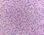
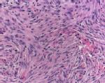
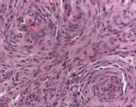
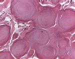
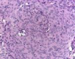
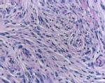
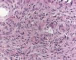
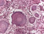
Meningiomas are tumours of the brain and nervous system. They account for 20% of all brain tumours. Mengiomas exist in three different grades of malignancy (WHO Grade I-III) most being benign (over 80%) but some showing an increased propensity to recurrence and rare cases being malignant. A meningioma at a later stage may develop into a mass of cells forming a protrusion against the brain that may cause damage to other brain cells due to limited space in the skull. It has a greater propensity of occurrence in an ageing population with very rare cases reported amongst children. Most benign WHO Grade I Meningiomas belong to one of the four subtypes, shown in the Figure below. The images were acquired by Mr Volkmar Hans at the Institute of Neuropathology, Bielefeld (Germany) using a Zeiss Axioskop 2 plus microscope fitted with a Zeiss Archoplan 40X 0,65 lens. As can be seen in the Figure, these images can have a high intra-class variation and low inter-class variation.
|
|
|
|
|
|
|
|
|
|
| Meningiotheliamatous | Fibroblastic | Transitional | Psammomatous |
In this project, we investigate methods for differentiating between the four subtypes of meningiomas. We have developed a robust and adaptive Wavelet Packets based representation that yields discriminant features for classification of a given meningioma image into one of the four subtypes.

Relevant Publications
- HA Qureshi, O Sertel, NM Rajpoot, RG Wilson, MN Gurcan,
Adaptive Discriminant Wavelet Packet Transform and Local Binary Patterns for Meningioma Subtype Classification,
Proceedings 11th Medical Image Computing and Computer-Assisted Intervention (MICCAI'2008), September 2008

- HA Qureshi, RG Wilson, NM Rajpoot,
Optimal Wavelet Basis for Wavelet Packets based Meningioma Subtype Classification,
Proceedings 12th Medical Image Understanding and Analysis (MIUA'2008), July 2008
- HA Qureshi, NM Rajpoot, RG Wilson, TW Nattkemper, V Hans,
Comparative Analysis of Discriminant Wavelet Packet Features and Raw Image Features for Classification of Meningioma Subtypes,
Proceedings Medical Image Understanding and Analysis (MIUA'2007), July 2007
- HA Qureshi, NM Rajpoot, K Masood, V Hans,
Classification of Meningiomas using Discriminant Wavelet Packets and Learning Vector Quantization,
Proceedings of Medical Image Understanding and Analysis (MIUA'2006), July 2006